Biography

Technical leader with 18 years of hands-on experience in medical imaging, artificial intelligence, computer vision, data science and neuroscience.

I work at the Scientific Computing and Imaging (SCI) Institute, as director of the Biomedical imaging, data science and AI core (BIDAC), a University of Utah health science service center. BIDAC offers application-oriented consulting services, developing and delivering novel AI solutions to medical device startups and research groups in healthcare, health sciences and material science. My role entails both business operations and technical expertise, helping partners turn data into insights.

Expertise

Technical skills

  • Medical imaging
  • Artificial intelligence
  • Computer vision
  • Deep learning
  • Machine learning
  • Data science
  • Generative AI

Business skills

  • Consulting
  • Account management
  • Project management
  • Product management
  • Budget management
  • Team leadership

Areas of Interests

  • Healthcare
  • Health sciences
  • Neuroscience
  • AI - Generative AI
  • Business applications
  • International relations

Project collaborations

I have been leading and developing end-to-end computer vision, AI and data science solutions, working in multi-institutional, interdisciplinary teams of computer scientists, software engineers, health science researchers and medical investigators.

From 2007 to 2012, I led the development of multiple open-source software products used in various neuroscience applications, within the multi-center National Alliance for Medical Image Computing. From 2012 to 2016, I designed and applied end-to-end image analysis solutions for the multi-hospital Autism Center of Excellence Network - Infant Brain Imaging Study, processing 1000+ imaging studies.

Since 2016 via BIDAC, I built various computer vision and AI solutions for both academic research groups and start-up companies. Multidisciplinary projects include AI-based 3D sensing for autonomous systems, image classification tasks for material science via deep learning, GPU computing for health sciences and AI-based 3D segmentation tasks for healthcare. Please find some highlights below.

Nuclear forensic analysis via deep learning

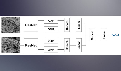
Nuclear forensics aims to investigate the origin and history of nuclear or radioactive materials via analytical techniques. Deep learning applied to electron microscopy images can automatically and accurately identify subtle differences in microstructure from different samples. It provides insights into synthesis conditions and processing routes of uranium ore concentrates. We applied, compared and fine-tuned via transfer learning state-of-the-art convolutional neural networks, performing numerous image classification and regression tasks. In addition to applying standard neural network architectures (ResNet, DenseNet), we implemented parallel networks to study multi-magnification acquisitions, and quantified model uncertainty during inference.

AI-based 3D depth sensing from thermal camera systems

Long-range 3D perception is an essential capability for autonomous navigation. The additional use of Long Wave Infrared (LWIR) cameras, compared to other systems, enable environment awareness in degraded visual conditions, such as fog, dust or low illumination. By leveraging a multi-sensor approach and the use of deep learning, we aim to reduce low contrast and low resolution often associated with thermal imaging, making it relevant for 3D sensing. We built multi-input single-output deep neural networks, enabling long-range 3D perception from stereo thermal camera systems. The combination of multiple sensors and AI-based analysis enabled accuracy improvement compared to simple stereo-vision and traditional image analysis methods.

Data engineering workflow for AI in radiology imaging

We aimed to investigate the use of machine learning on large retrospective radiology imaging studies, including chest x-rays and related clinical text reports. In partnership with researchers from the Radiology Department, the Enterprise Data Warehouse and the Center for High Performance Computing, we leveraged various software and hardware infrastructure to enable secured data transfer from the hospital PACS system, HIPAA-compliant data storage via a database, and data management of large radiological datasets. We retrieved data from 59,000+ patients (140,000+ studies), to study chronic obstructive pulmonary disease (COPD), COVID-19 and acute respiratory infections.

Computer vision for neuroscience studies (12+ years)

Over 12 years, I led and executed parallel imaging data science projects to support various national clinical research studies. These entailed automated image registration, atlas generation, tissue classification, structural segmentation and DTI analysis from multi-modal acquisitions. Studies focused on rodents, pigs and mostly human subjects ranging from neonatal age to elderly. Driving clinical applications included brain development in autism spectrum disorder, Parkinson's disease, Down syndrome, Huntington's disease, deep brain stimulation, and obsessive-compulsive disorder.