Pictures taken from demos and data sets of the
ANNOT3D PACKAGE
Poster Images:- MMVR Poster
- EngDay Poster
Bo's Bones:- Scapula
- Clavicle
- Humerus
- 3ShoulderBones
Head Demo:- ear picked
- plane cut
- sphere cut
- cube cut
Mummy Example:- MummyBones
Smile Example:- SmilePhantom
Other Images:- Miscellaneous
Poster Images
Medicine Meets Virtual Reality - Jan 15-17 2004, Newport Beach, CA
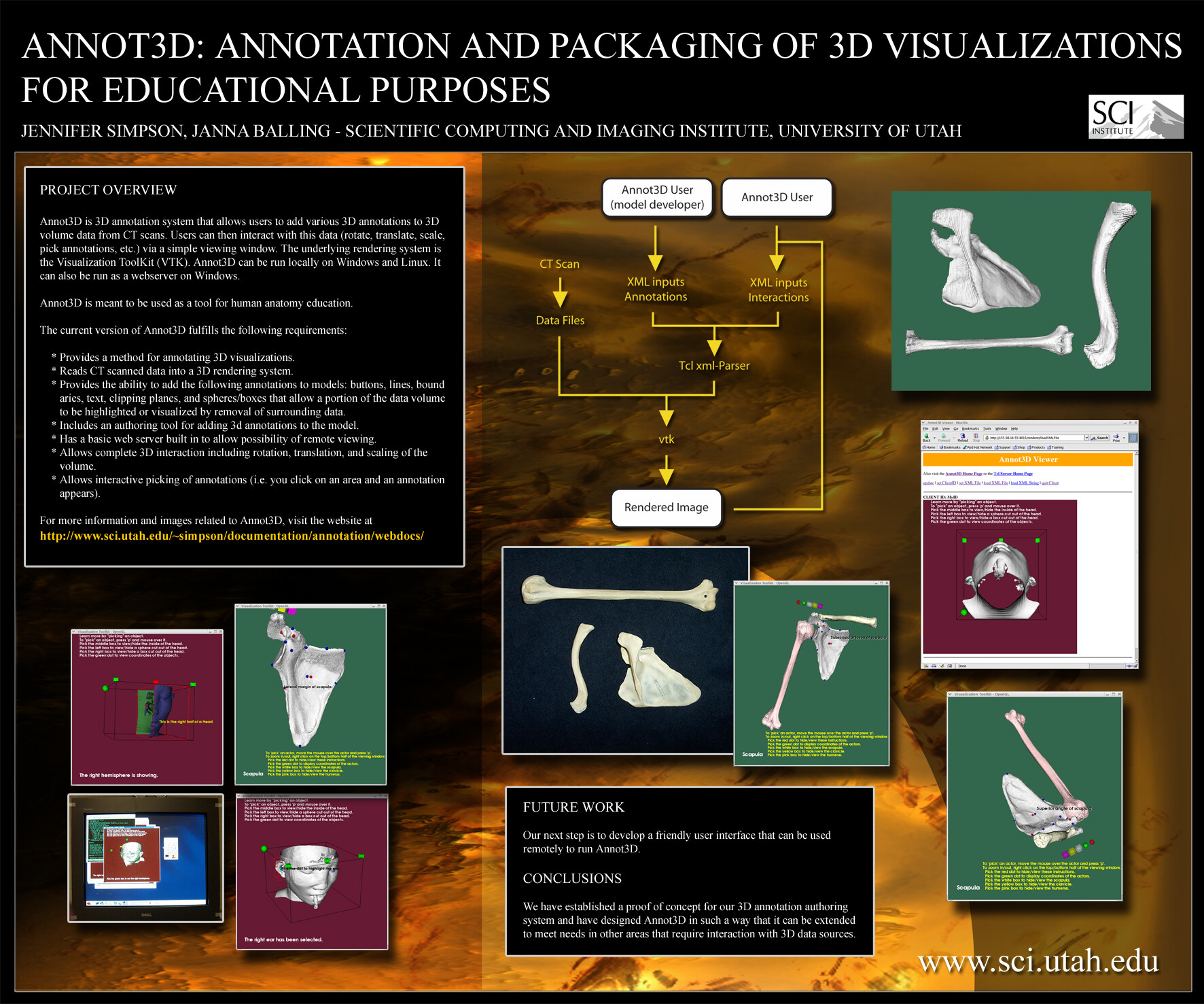
ANNOT3D: ANNOTATION AND PACKAGING OF 3D VISUALIZATIONS FOR EDUCATINAL PURPOSES
JENNIFER SIMPSON, JANNA BALLING - SCIENTIFIC COMPUTING AND IMAGING INSTITUTE, UNIVERSITY OF UTAH
PROBLEMA current challenge in university level anatomy education is the creation and distribution of accurate 3D anatomical visualizations to students for review and self-guided study. While many tools exist that provide pre-annotated 3D atlases, there is a lack of software that provides instructors and other users the ability to add customized annotations to their own digitally scanned 3D data.
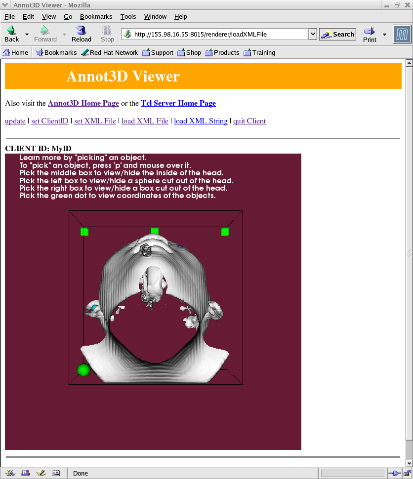
PROJECT OVERVIEWAnnot3D is 3D annotation system that allows users to add various 3D annotations to 3D volume data from CT scans. Users can then interact with this data (rotate, translate, scale, pick annotations, etc.) via a simple viewing window. The underlying rendering system is the Visualization ToolKit (VTK). Annot3D can be run locally on Windows and Linux. It can also be run as a webserver on Windows, although this feature needs more development before it is deployed to users.
Annot3D is meant to be used as a tool for human anatomy education.
The current version of Annot3D fulfills the following requirements:
For more information and images related to Annot3D, visit the website at http://www.sci.utah.edu/~simpson/documentation/projects/annotation/webdocs/
- Provides a method for annotating 3D visualizations.
- Reads CT scanned data into a 3D rendering system.
- Provides the ability to add the following annotations to models: buttons, lines, boundaries, text, clipping planes, and spheres/boxes that allow a portion of the data volume to be highlighted or visualized by removal of surrounding data.
- Includes an authoring tool for adding 3d annotations to the model. The Annot3D authoring system currently consists of XML files generated by hand to contain all information related to a dataset and its annotations.
- Has a basic web server built in to allow possibility of remote viewing.
- Allows complete 3D interaction including rotation, translation, and scaling of the volume.
- Allows interactive picking of annotations (i.e. you click on an area and an annotation appears).
Cutting plane annotation on a sample dataset, enabling users to view and annotate interior regions of interest. "Highlight" annotation designates a region of interest. RESULTS
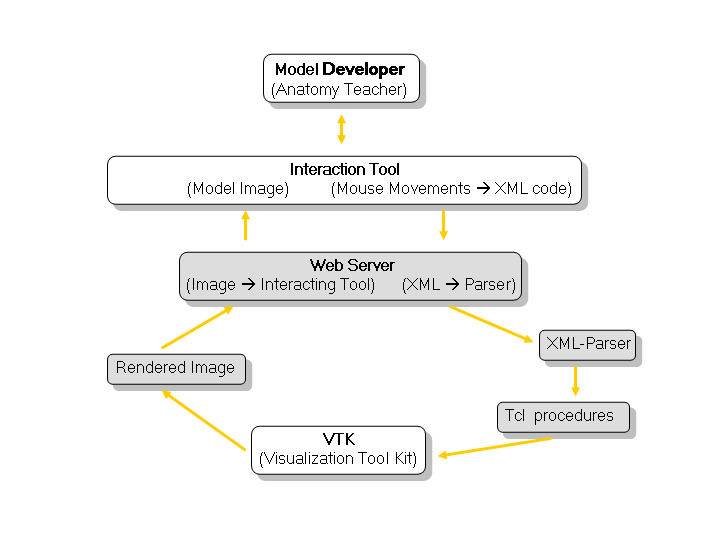
This diagram represents the flow of data and control in the Annot3D software system. The images surrounding the diagram are snapshots of various points within the flow of the system.We have produced a working implementation of our 3D annotation and authoring system. We have designed Annot3D in such a way that it can be extended to meet needs in other areas that require interaction with 3D data sources.
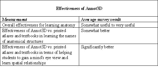
CONCLUSIONBased on our usability studies we found that Annot3D is a useful tool for helping students to learn anatomy and spacial relationships. There is still much work to be done to make Annot3D user-friendly enough for the average anatomy student or instructor. This work will be part of future phases of the project.
FUTURE WORK
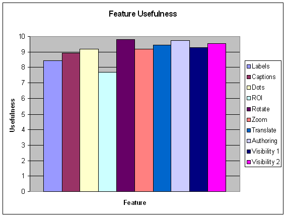
Results from a survey given to eleven medical students regarding the effectiveness of Annot3D as a learning tool for anatomy education. The students were asked to rate the usefulness of each feature on a scale of 1 to 10. Results from a survey given to eleven medical students regarding the usefulness of various Annot3D features. Our next step is to incorporate user feedback to develop a friendly user interface that can be used locally or possibly remotely to run Annot3D.
This work was supported by the National Science Foundation (grant #ACI-9619019 PI-Chris Johnson, Ph.D.)
Engineering Day - Fall 2003
Bo's Bones
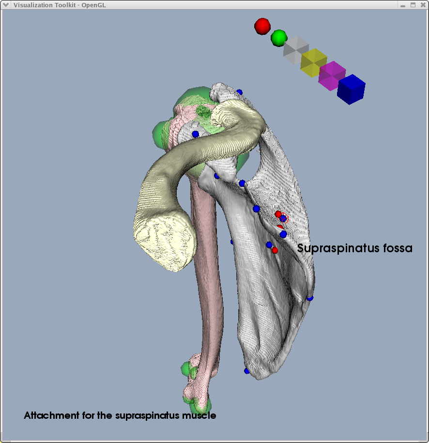
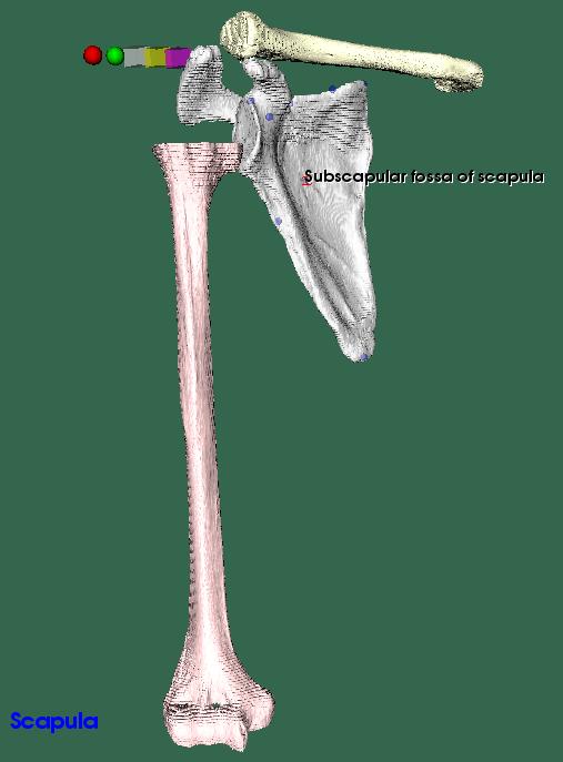
Scapula

This is a picture of the Scapula. Taken from Bo's CT data scanned at 1.3 mm resolution. This has an isosurface value of 100.02
Clavicle

This is a picture of the Clavicle. Taken from Bo's CT data scanned at 1.3 mm resolution. This has an isosurface value of 100.02
Humerus

This is a picture of the Humerus. Taken from Bo's CT data scanned at 1.3 mm resolution. This has an isosurface value of 100.03
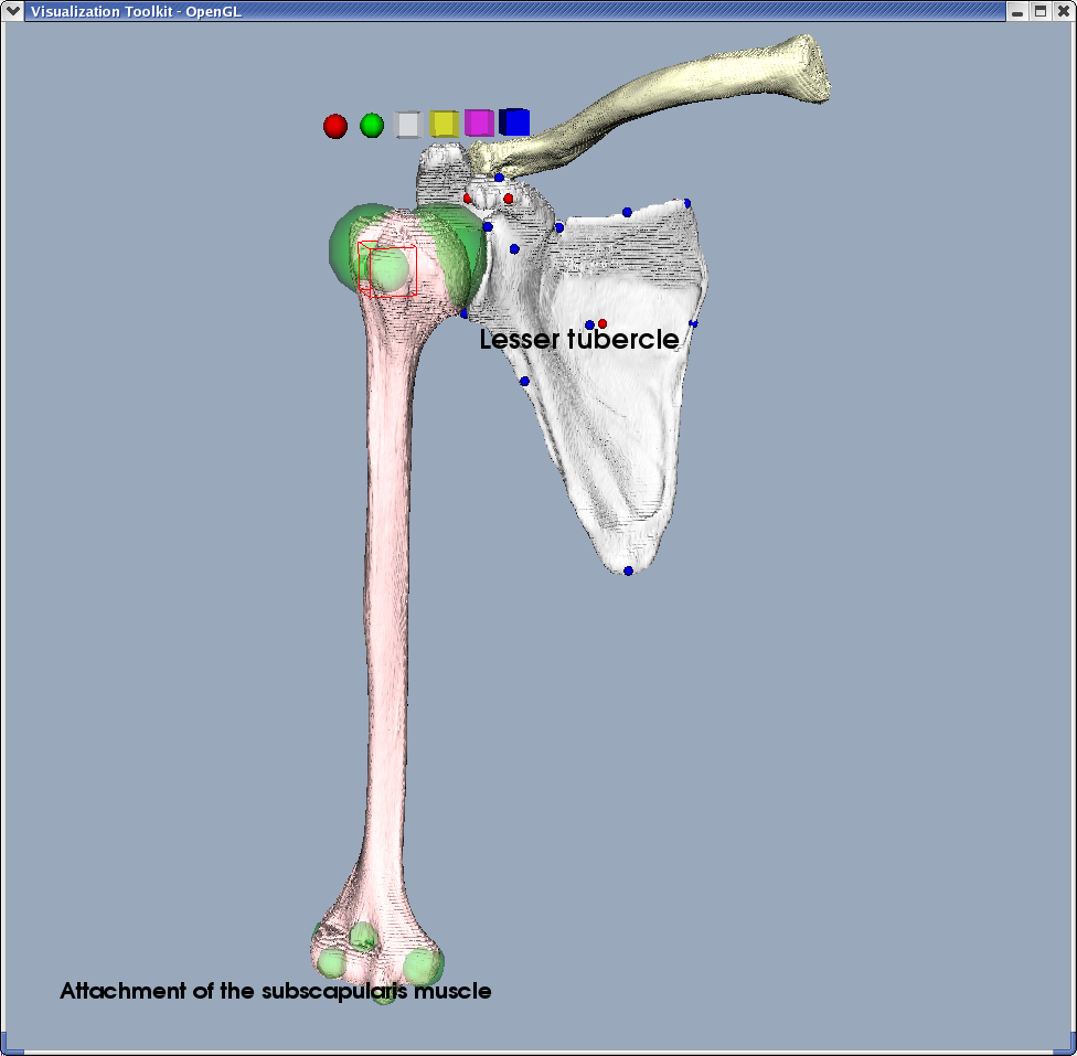
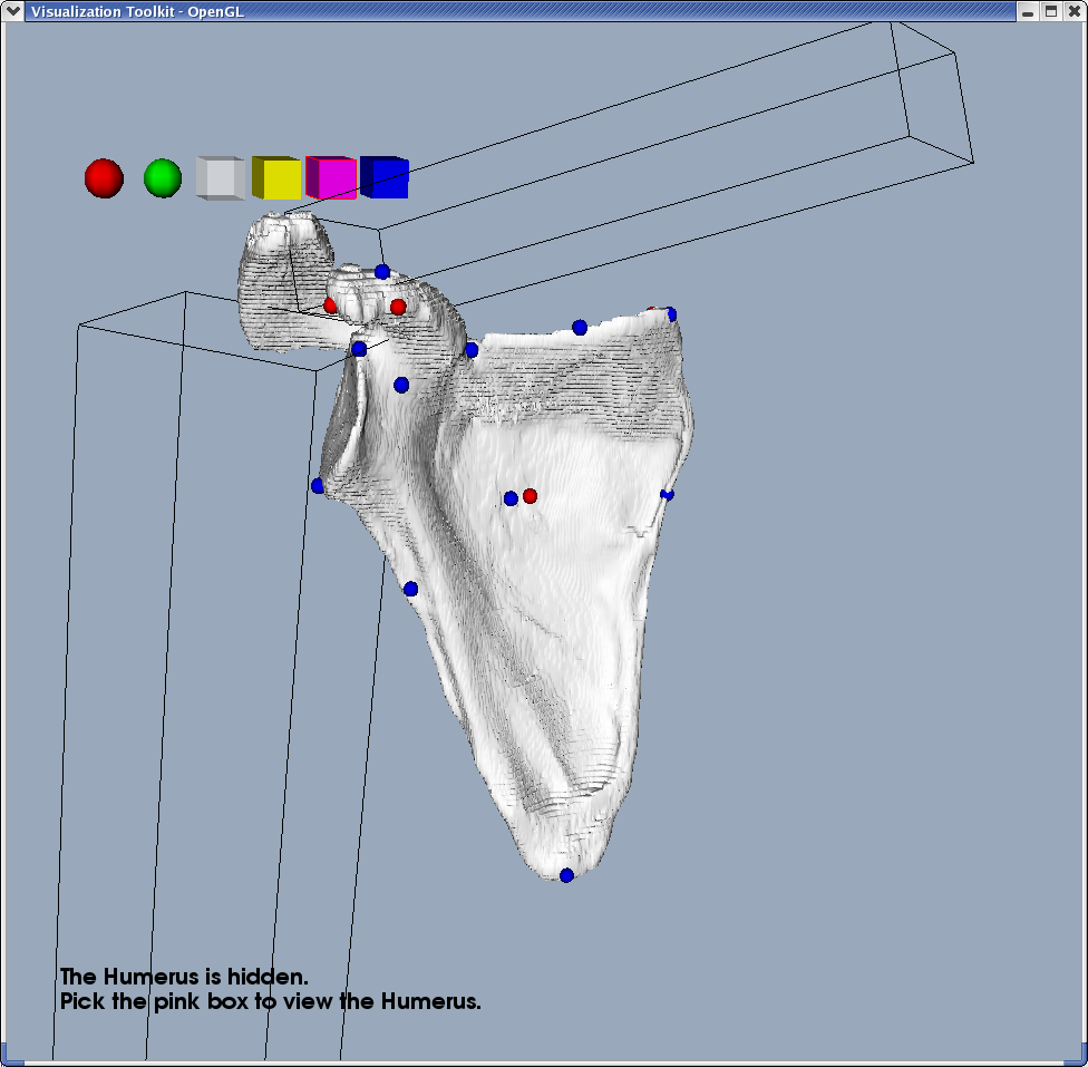
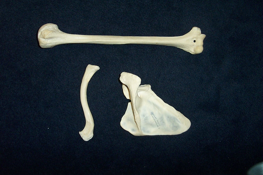
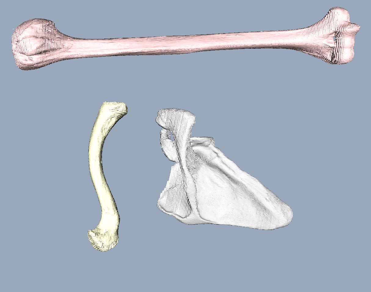
3ShoulderBones
This is a picture of the Scapula, Clavicle, and Humerus. Taken from Bo's CT data scanned at 1.3 mm resolution. These do have anatomically correct orientation, but the relative scale of the three bones is slightly off (the clavicle needs to be smaller).
Head Demo
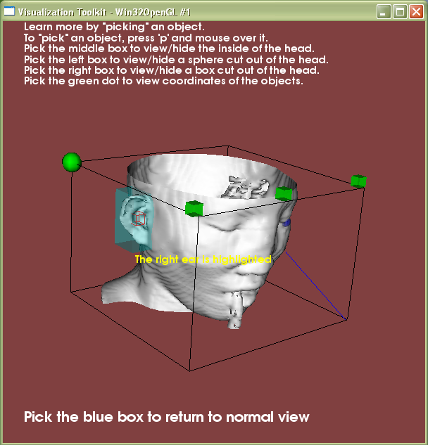
HeadDemo - ear picked
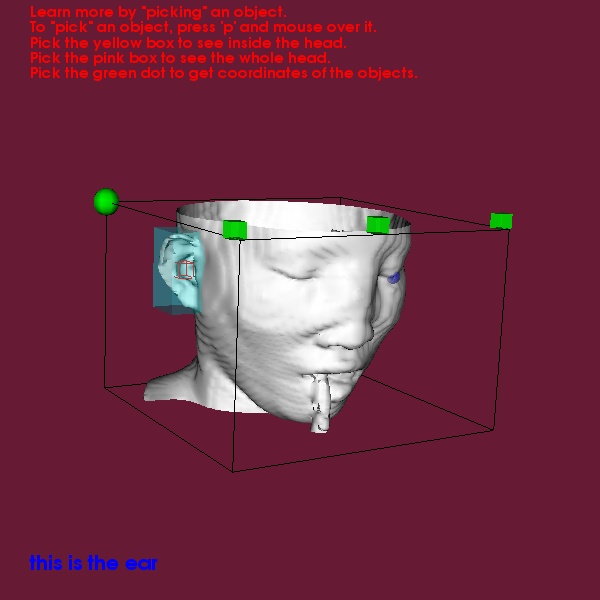
This is a picture of the HeadDemo with a highlighted ear. Taken from data in the vtk data examples directory.
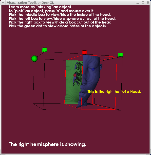
HeadDemo - plane cut

This is a picture of the HeadDemo with a clipping plane. Taken from data in the vtk data examples directory.
HeadDemo - sphere cut
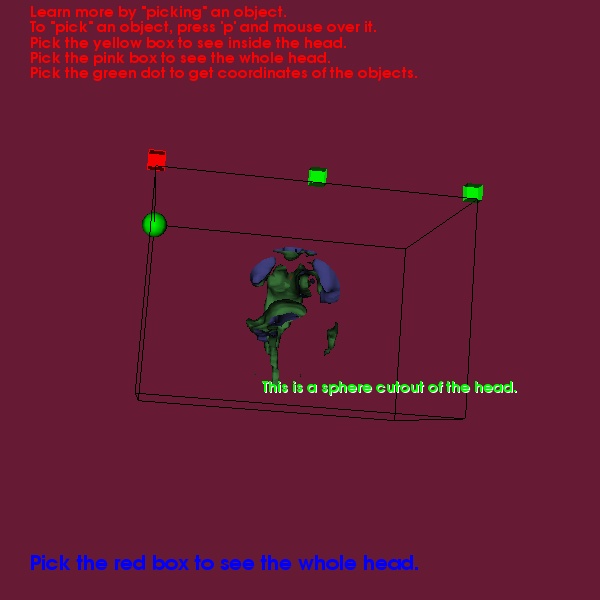
This is a picture of the HeadDemo with a clipping sphere. Taken from data in the vtk data examples directory.
HeadDemo - cube cut
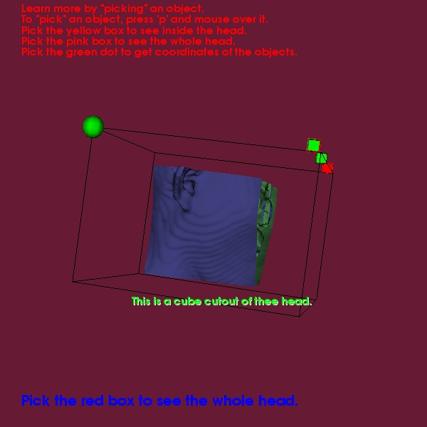
This is a picture of the HeadDemo with a clipping cube. Taken from data in the vtk data examples directory.
Mummy Example
MummyBones
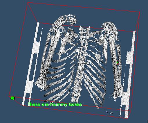
This is a picture of the mummyExample bones. Taken from CT data in Greg Jones' collection.
Smile Example
SmilePhantom
This is a picture of the SmileExample CT phantom. Taken from sample CT data on the dicom web site.
Other Images
Miscellaneous
view folder contents