 |
The final class of validation methods we will discuss manages to circumvent the limitations of human-based measurements by identifying clinical diagnoses or other a priori clinical information that can serve as (sometimes indirect) measures of electrocardiographic variables. The first requirement of validation in inverse electrocardiography, obtaining accurate geometric information, is possible through the use of modern medical imaging. Magnetic resonance (MR) and computed tomography (CT) are the two complementary techniques that provide the required spatial resolution as well as a means of identifying inhomogeneous regions (soft tissue from MR and bone from CT) from variation in image intensity. An important requirement of some medical imaging modalities for cardiac applications is the need to time the acquisition of data with the cardiac cycle. Temporal resolution of MR imaging, for example, is not yet adequate to sample the entire heart region within the diastolic interval of the heart so that multiple images gated to the ECG must be sampled and combined. A full gated MR tomography of the thorax can take many minutes to complete so that variations in cardiac shape can lead to errors in the resulting geometric models. Chest movement associated with breathing is another source of error in ECG-gated scans because the respiration cycle is independent of heart rhythm even as it couples via the diaphragm and lungs to alter heart position (see, for example, Pflugfelder[91]). The acquisition of accurate patient geometry remains a significant obstacle to application of inverse solutions to clinical settings. One consequence is the intense research interest in determining how dependent inverse solution results are on geometric accuracy, and how best to approximate that geometry with less time, effort, and cost.[10,28,92,93,94]
Even with good quality tomographic images, the conversion to discrete geometric models is not a trivial undertaking. The first step is to identify contours that mark the boundaries between regions of interest, known as ``segmentation'', a subject which is the topic of ongoing research (see, for example, Hinshaw et al.[95] or Calabi et al.[96]) and for which no reliable completely automatic scheme exists. From these contours, it is then possible to construct triangular surface meshes[97,98,99,100] or volume meshes from tetrahedra[101,102,103,104,105,94] and hexahedra.[106] At present, construction of a detailed human geometric model still requires considerable technical expertise and time but is a feasible undertaking.
Obviously, the real difficulty with clinical validation lies in the second requirement--direct measurement of cardiac sources at an adequate spatial resolution is usually impossible, especially under closed-chest conditions. Some investigators have carried out inverse solutions and compared the signals at selected epicardial sites to measured electrograms from individual electrodes implanted chronically in patients.[106,107] Budgett et al. recorded from six electrodes attached to the epicardium during coronary bypass surgery and compared the signals to those estimated over patches extracted from a finite difference calculation based on customized torso geometries.[106] They found reasonable agreement of signal morphology (but not amplitude) in four of the six sites, but had to time shift the signals by as much as 26 ms to achieve correlation levels in the range of 0.91-0.98. Two of the sites showed very little agreement between measurements and calculations.
Another approach to obtaining source data in patients is to apply epicardial sock electrodes during open chest surgery. Shahidi et al. performed a study in which they created patient specific geometric models and then recorded body surface potentials before the surgery.[20] They compared computed epicardial potential maps with those measured during surgery and found only approximate agreement. The main problem with the general approach of measuring body surface maps at one time and epicardial potentials during open chest surgery at a different time is that the effects of the resulting changes in patient state and the integrity of the volume conductor are poorly known. There is so much variability introduced by these disparate measurement conditions that it is virtually impossible to decide how to separate the resulting errors between the inverse solution and the measurements.
In lieu of direct validation, inverse solutions can sometimes be evaluated based on our general knowledge of what constitutes a reasonable description of, for example, the activation sequence. Huiskamp et al. performed inverse calculations of the epicardial and endocardial activation sequence[27,28] and compared their results to the seminal publication by Durrer et al. on the measured activation sequence of an isolated, healthy human heart.[19] They also used another indirect validation measure by first estimating the activation time from the body surface potentials and then computing in a forward sense the body surface ECGs, against which they could compare original measurements.[27,28] Budget et al. also compared their computed epicardial potentials against those described by Spach et al. for chimpanzee hearts[108,109] as a means of validation.[106]
Clinical conditions can facilitate validation of inverse solutions by providing non-electrocardiographic indicators of disease states that are also revealed by epicardial potentials. Kilpatrick et al. described an early example of this approach in which they used computed epicardial potential distributions from the ST segment of the ECG to predict the vessel affected in cases of acute myocardial infarction.[1] To confirm the location of infarction, they performed angiography on each patient in the study and then quantified their results in terms of correct or incorrect prediction of the affected vessel. In a subsequent study Kilpatrick et al. used inverse solutions of epicardial potentials to differentiate the cardiac origins of ST-segment depressions visible in the body surface.[110]
Another example of a clinical condition for validation is Wolff-Parkinson-White (WPW) syndrome, in which an accessory conductive pathway exists between the atria and the ventricles, which are normally electrically isolated.[111] The advantage of this condition from the perspective of the study of inverse solutions is that when the accessory path is active, there is a discrete deflection in the body surface ECG known as the ``delta wave'', which occurs just before the onset of the R wave. It is possible to apply inverse solutions to a few time instants during the delta wave and attempt to locate the accessory pathway. The very local extent of active tissue during the delta wave also justifies the use of dipole sources and thus inverse solutions based on epicardial surface potentials and activation times as well as discrete source models. Confirmation of the predicted accessory pathway site is then possible by means of either open chest cardiac mapping[112] or by catheter techniques in common use.[113] Shahidi et al.[20] and Penney et al.[114] in separate studies applied inverse solutions to WPW patients and compared the predicted accessory pathway with that found subsequently during ablation.
One limitation of all the clinical approaches described so far is the lack of control over the source potentials. Patients either have or do not have a particular clinical abnormality and it is often difficult or impossible to alter that state. Patients with WPW syndrome are one exception in that it is often possible to illicit both normal sinus beats and beats with pre-excitation via the accessory pathway during the same electrophysiology study. There is, however, a palliative intervention that allows direct and continuous control over some aspects of the electrophysiologic state of the heart and thus lends itself to collateral use for validation of inverse solutions. During percutaneous transluminal coronary angioplasty (PTCA), a catheter balloon is inserted into a partially occluded coronary artery and inflated in order to mechanically disrupt the thickened and hardened linings of the artery.[115] During the inflation, which typically lasts from 20-200 s, blood flow in the affected artery is completely occluded and acute ischemia results. Electrophysiologic consequences of this ischemia are visible from the body surface ECG and resolve within seconds to minutes of the balloon deflating. The inflation/deflation cycle can be--and often is for clinical reasons--repeated many times in the same patient, both in the same and different segments of the coronary arteries.
PTCA has served as a human model of many aspects of cardiac physiology during acute ischemia[116] including ventricular contraction,[117] wall thickening,[118] coronary hemodynamics,[119] collateral flow,[120] left ventricular filling,[121] and the genesis of arrhythmias.[122] MacLeod et al. used PTCA to validate an electrocardiographic inverse solution.[18,2] They computed epicardial potentials from body surface maps of patients during angioplasty and compared the predicted locations of epicardial ST-segment elevations with the locations of the balloon catheter documented via angiography. Working mostly with time-integrated potentials, they found patterns of torso potentials during inflation that were characteristic of the vessel under treatment. When used as inputs to inverse solutions, these torso potentials generated associated epicardial distributions that were similar to ST-segment potential changes seen in animal models of acute ischemia. Thus, in an optimistic interpretation, these inverse solutions may have succeeded in localizing focused areas of change in epicardial potential due to the induced ischemia from a diffuse change observed on the body surface. In fact, in many cases, elevations in ST-segment potentials on the epicardium corresponded to locations just distal to the site of the angioplasty balloon, within what could reasonably be assumed to be the approximate perfusion area supplied by the distal circumflex artery.
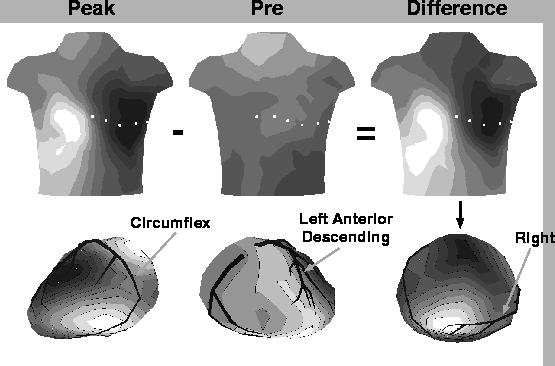
Figure 4 shows examples of body surface maps and computed inverse solution from angioplasty patients. The top row of images highlights a particularly powerful feature of the PTCA validation model, which is the ability to apply and measure the effects of controlled interventions. The leftmost body surface maps shows the isointegral distribution of the ST segment during the last 30 s of an inflation lasting 150 s and the middle map shows the equivalent isointegral map recorded just prior to the onset of balloon inflation. The rightmost map is the difference between the other two (peak- minus pre-inflation) and shows clearly the electrophysiologic influence of the transient occlusion on the body surface potentials. The light shaded (positive) area on the right anterior chest in the left and right maps is a typical characteristic of at least some of the patients undergoing angioplasty of the right coronary (RC) artery. The rightmost epicardial map in the bottom row of the figure contains the inverse computed isointegral distribution from the torso difference map directly above. The light area in this map lies over the terminal region of the RC artery and suggests that PTCA induced an elevation of ST-segment potentials during the balloon inflation, in agreement with the angiographic evidence for this patient. The left and middle epicardial maps in the lower row of the figure are inverse solutions computed from ST-segment isointegral difference maps from two other patients undergoing circumflex and left anterior descending artery PTCA, respectively. Here, too, the light shaded region indicates a positive potential change that lies over the region of expected underperfusion, again suggesting that one can detect locations of ischemic myocardium by means of an inverse solution.
|
While these validation results were encouraging, they were still very qualitative in nature, and contained unexplained anomalies such as secondary areas of positive potential in the atrial region or in parts of the ventricle where there was no angiographic evidence to indicate ischemia. The geometry used in this study was realistic but not anatomically accurate nor did it contain inhomogeneous regions. The heart was based on a detailed model created from a human heart and used for cellular automata simulations of propagation[41,123,124] and it was placed in the torso model from a different subject created by mechanical measurements.[2,125,126]
A recent report by Tilg et al. described a validation approach in humans that offers exceptional potential for testing at least one form of the inverse solution.[127] The breakthrough technology in this study was the use of a remote ranging system that measures activation time and location simultaneously from an endocardial catheter.[128] Tilg et al. recorded from a series of patients the endocardial activation times as well as simultaneous body surface potentials. They then performed MR imaging and a magnetocardiographic mapping, all within a day of the endocardial mapping. Using the geometry and the body surface potentials, they then created customized inverse solutions in terms of activation time, which they could validate against the direct measurements. Preliminary results suggest a reasonably good match between predicted and measured values.
It is also possible to formulate an inverse solution in terms of reconstructing endocardial potentials from potentials measured in the interior of the cavities in the heart. Such an approach has been developed by Khoury et al.[80,129] and Lui et al..[82] Initial validation of this approach was by means of animal studies but recently this group has developed a combined probe that contains both a set of non-contact electrodes mounted on a cylinder, and a concentric basket catheter.[130] In this way, it is possible to record from both electrode arrays simultaneously and directly validate the computed endocardial potentials against measurements.