
The SCI Head Model is free and open to the public. All we ask is that appropriate acknowledgements be included in published works when using this data repository. The accompanying paper: “A High-Resolution Head and Brain Computer Model for Forward and Inverse EEG Simulation” contains specific instructions on how to build and use this model specifically for forward in inverse EEG simulations.
If you have any questions please email: .
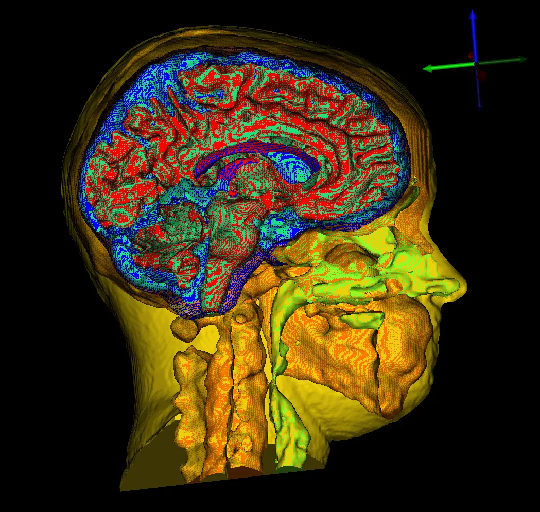
Imaging and electroencephalography for this project were done at Electrical Geodesics Inc. at the University of Oregon. The SCI Head Model is an open-source data repository that is principally funded through the SCI Institute’s NIH/NIGMS Center for Integrative Biomedical Computing (CIBC). Please use the following acknowledgments and send us references to any publications, presentations, or successful funding applications that make use of NIH/NIGMS CIBC software or data sets.
“This project was supported by the National Institute of General Medical Sciences of the National Institutes of Health under grant numbers P41 GM103545 and R24 GM136986.”
A. Warner, J. Tate, B. Burton, and C.R. Johnson. 2019. A High-Resolution Head and Brain Computer Model for Forward and Inverse EEG Simulation. bioRxiv doi: 10.1101/552190.
Acquisition Reports:
All acquisition reports for T1-weighted and T2-weighted magnetic resonance imaging (MRI), diffusion-weighted imaging (DWI), functional MRI (fMRI), and electroencephalogram (EEG) recordings. Imaging and electroencephalography for this project were done at Electrical Geodesics Inc. at the University of Oregon.