


SCI Publications
2006


L. Barbosa, J. Freire.
“Automatically Constructing Collections of Online Databases,” In Proceedings of the 15th ACM International Conference on Information and Knowledge Management (CIKM), Arlington, pp. 796--797. 2006.
S. Basu, P.T. Fletcher, R.T. Whitaker.
“Rician Noise Removal in Diffusion Tensor MRI,” In Medical Image Computing and Computer Assisted Intervention (MICCAI), Vol. LNCS 4190, pp. 117--125. October, 2006.
L. Bavoil, S.P. Callahan, C.T. Silva.
“Robust Soft Shadow Mapping with Depth Peeling,” SCI Institute Technical Report, No. UUSCI-2006-028, University of Utah, 2006.
L. Bavoil, S.P. Callahan, A. Lefohn, J.L.D. Comba, C.T. Silva.
“Multi-Fragment Effects on the GPU using the k-Buffer,” SCI Institute Technical Report, No. UUSCI-2006-032, University of Utah, 2006.
“Advances in Visual Computing, Second International Symposium, ISVC 2006, Lake Tahoe, NV, USA, November 6-8, 2006 Proceedings, Part I,” In ISVC (1), Lecture Notes in Computer Science, Vol. 4291, Edited by George Bebis and Richard Boyle and Bahram Parvin and Darko Koracin and Paolo Remagnino and Ara V. Nefian and Meenakshisundaram Gopi and Valerio Pascucci and Jiri Zara and Jose Molineros and Holger Theisel and Thomas Malzbender, Springer, 2006.
ISBN: 3-540-48628-3
C. Benthin, I. Wald, P. Slusallek.
“Techniques for Interactive Ray Tracing of Bezier Surfaces,” In Journal of Graphics Tools, Vol. 11, No. 2, pp. 1--16. 2006.
C. Benthin, I. Wald, M. Scherbaum, H. Friedrich.
“Ray Tracing on the CELL Processor,” In Proceedings of the 2006 IEEE Symposium on Interactive Ray Tracing, pp. 25--32. 2006.
F.F. Bernardon, S.P. Callahan, J.L.D. Comba, C.T. Silva.
“Interactive Volume Rendering of Unstructured Grids with Time-Varying Scalar Fields,” In Proceedings of the Eurographics Symposium on Parallel Graphics and Visualization, pp. 51--58. 2006.
M. Berzins.
“Is there Still More to Science than Simulation?,” No. UUSCI-2006-031, SCI Institute, University of Utah, November, 2006.
M. Berzins.
“Adaptive Polynomial Interpolation on Evenly Spaced Meshes,” SCI Institute Technical Report, No. UUSCI-2006-033, University of Utah, 2006.
W. Bethel, C.R. Johnson, C.D. Hansen, S.G. Parker, A.R. Sanderson, C.T. Silva, X. Tricoche, V. Pascucci, H. Childs, J. Cohen, M. Duchaineau, D. Laney, P. Lindstrom, S. Ahern, J. Meredith, G. Ostrouchov, K. Joy, B. Hamann.
“VACET: Proposed SciDAC2 Visualization and Analytics Center for Enabling Technologies,” In J. Phys.: Conf. Ser., Vol. 46, pp. 561--569. 2006.
J. Bigler, A.J. Stephens, S.G. Parker.
“Design for Parallel Interactive Ray Tracing Systems,” SCI Institute Technical Report, No. UUSCI-2006-027, University of Utah, 2006.
J. Bigler, J. Guilkey, C. Gribble, C.D. Hansen, S.G. Parker.
“A Case Study: Visualizing Material Point Method Data,” In Proceedings of Euro Vis 2006, pp. 299--306, 377. May, 2006.
J. Bigler, A.J. Stephens, S.G. Parker.
“Design for Parallel Interactive Ray Tracing Systems,” In Proceedings of The IEEE Symposium on Interactive Ray Tracing, pp. 187--196. 2006.
S. Boulos, D. Edwards, J.D. Lacewell, J.M. Kniss, J. Kautz, P. Shirley, I. Wald.
“Interactive Distribution Ray Tracing,” SCI Institute Technical Report, No. UUSCI-2006-022, University of Utah, 2006.
S. Boulos, I. Wald, P. Shirley.
“Geometric and Arithmetic Culling Methods for Entire Ray Packets,” School of Computing Technical Report, No. UUCS-06-010, School of Computing, University of Utah, 2006.
P.-T. Bremer, W. Cabot, A. Cook, D. Laney, A. Mascarenhas, P. Miller, V. Pascucci.
“Understanding the Structure of the Turbulent Mixing Layer in Hydrodynamic Instabilities,” In Proceedings of SciDAC 2006 -- Scientific Discovery Through Advanced Computing, Denver, CO, USA, Vol. 46, Journal of Physics Conference Series, pp. 556--560. June, 2006.
S. Browd, L.J. Healy, G. Dobie, J.T. Johnson III, G.M. Jones, L.F. Rodriguez, D.L. Brockmeyer.
“Morphometric and Qualitative Analysis of Congenital Occipitocervical Instability in Children: Implications for Down Syndrome Patients,” In Journal of Neurosurgery: Pediatrics, Vol. 105, No. 1 , Journal of Neurosurgery Publishing Group, pp. 50--54. July, 2006.
DOI: 10.3171/ped.2006.105.1.50
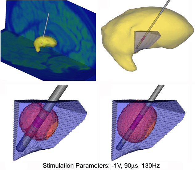
C.R. Butson, C.B. Maks, C.C. McIntyre.
“Sources and effects of electrode impedance during deep brain stimulation,” In Clinical Neurophysiology, Vol. 117, No. 2, pp. 447--454. 2006.
DOI: 10.1016/j.clinph.2005.10.007
PubMed ID: 16376143
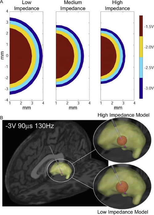
METHODS: Axisymmetric finite-element models (FEM) of the DBS system were constructed with explicit representation of encapsulation layers around the electrode and implanted pulse generator. Impedance was calculated by dividing the stimulation voltage by the integrated current density along the active electrode contact. The models utilized a Fourier FEM solver that accounted for the capacitive components of the electrode-tissue interface during voltage-controlled stimulation. The resulting time- and space-dependent voltage waveforms generated in the tissue medium were superimposed onto cable model axons to calculate the VTA.
RESULTS: The primary determinants of electrode impedance were the thickness and conductivity of the encapsulation layer around the electrode contact and the conductivity of the bulk tissue medium. The difference in the VTA between our low (790 Omega) and high (1244 Omega) impedance models with typical DBS settings (-3 V, 90 mus, 130 Hz pulse train) was 121 mm3, representing a 52\% volume reduction. CONCLUSIONS: Electrode impedance has a substantial effect on the VTA and accurate representation of electrode impedance should be an explicit component of computational models of voltage-controlled DBS.
SIGNIFICANCE: Impedance is often used to identify broken leads (for values > 2000 Omega) or short circuits in the hardware (for values during DBS.
Keywords: Brain, Brain: physiology, Computer Simulation, Deep Brain Stimulation, Electric Conductivity, Electric Impedance, Electrodes, Humans, Imaging, Models, Neurological, Three-Dimensional
C.R. Butson, C.C. McIntyre.
“Role of electrode design on the volume of tissue activated during deep brain stimulation,” In Journal of Neural Engineering, Vol. 3, No. 1, pp. 1--8. March, 2006.
ISSN: 1741-2560
DOI: 10.1088/1741-2560/3/1/001
PubMed ID: 16510937
Keywords: Animals, Brain, Brain: physiology, Computer Simulation, Computer-Aided Design, Deep Brain Stimulation, Deep Brain Stimulation: instrumentation, Deep Brain Stimulation: methods, Electrodes, Equipment Design, Equipment Design: methods, Equipment Failure Analysis, Equipment Failure Analysis: methods, Humans, Implanted, Microelectrodes, Models, Neurological, Neurons, Neurons: physiology, Organ Size, Organ Size: physiology