


SCI Publications
2022


C. A. Nizinski, C. Ly, C. Vachet, A. Hagen, T. Tasdizen, L. W. McDonald.
“Characterization of uncertainties and model generalizability for convolutional neural network predictions of uranium ore concentrate morphology,” In Chemometrics and Intelligent Laboratory Systems, Vol. 225, Elsevier, pp. 104556. 2022.
ISSN: 0169-7439
DOI: https://doi.org/10.1016/j.chemolab.2022.104556
As the capabilities of convolutional neural networks (CNNs) for image classification tasks have advanced, interest in applying deep learning techniques for determining the natural and anthropogenic origins of uranium ore concentrates (UOCs) and other unknown nuclear materials by their surface morphology characteristics has grown. But before CNNs can join the nuclear forensics toolbox along more traditional analytical techniques – such as scanning electron microscopy (SEM), X-ray diffractometry, mass spectrometry, radiation counting, and any number of spectroscopic methods – a deeper understanding of “black box” image classification will be required. This paper explores uncertainty quantification for convolutional neural networks and their ability to generalize to out-of-distribution (OOD) image data sets. For prediction uncertainty, Monte Carlo (MC) dropout and random image crops as variational inference techniques are implemented and characterized. Convolutional neural networks and classifiers using image features from unsupervised vector-quantized variational autoencoders (VQ-VAE) are trained using SEM images of pure, unaged, unmixed uranium ore concentrates considered “unperturbed.” OOD data sets are developed containing perturbations from the training data with respect to the chemical and physical properties of the UOCs or data collection parameters; predictions made on the perturbation sets identify where significant shortcomings exist in the current training data and techniques used to develop models for classifying uranium process history, and provides valuable insights into how datasets and classification models can be improved for better generalizability to out-of-distribution examples.
2021
C. Ly, C. Nizinski, C. Vachet, L. McDonald, T. Tasdizen.
“Learning to Estimate the Composition of a Mixture with Synthetic Data,” In Microscopy and Microanalysis, 2021.
Identifying the precise composition of a mixed material is important in various applications. For instance, in nuclear forensics analysis, knowing the process history of unknown or illicitly trafficked nuclear materials when they are discovered is desirable to prevent future losses or theft of material from the processing facilities. Motivated by this open problem, we describe a novel machine learning approach to determine the composition of a mixture from SEM images. In machine learning, the training data distribution should reflect the distribution of the data the model is expected to make predictions for, which can pose a hurdle. However, a key advantage of our proposed framework is that it requires reference images of pure material samples only. Removing the need for reference samples of various mixed material compositions reduces the time and monetary cost associated with reference sample preparation and imaging. Moreover, our proposed framework can determine the composition of a mixture composed of chemically similar materials, whereas other elemental analysis tools such as powder X-ray diffraction (p-XRD) have trouble doing so. For example, p-XRD is unable to discern mixtures composed of triuranium octoxide (U3O8) synthesized from different synthetic routes such as uranyl peroxide (UO4) and ammonium diuranate (ADU) [1]. In contrast, our proposed framework can easily determine the composition of uranium oxides mixture synthesized from different synthetic routes, as we illustrate in the experiments.
C. Ly, C. A. Nizinski, A. Toydemir, C. Vachet, L. W. McDonald, T. Tasdizen.
“Determining the Composition of a Mixed Material with Synthetic Data,” In Microscopy and Microanalysis, Cambridge University Press, pp. 1--11. 2021.
DOI: 10.1017/S1431927621012915
Determining the composition of a mixed material is an open problem that has attracted the interest of researchers in many fields. In our recent work, we proposed a novel approach to determine the composition of a mixed material using convolutional neural networks (CNNs). In machine learning, a model “learns” a specific task for which it is designed through data. Hence, obtaining a dataset of mixed materials is required to develop CNNs for the task of estimating the composition. However, the proposed method instead creates the synthetic data of mixed materials generated from using only images of pure materials present in those mixtures. Thus, it eliminates the prohibitive cost and tedious process of collecting images of mixed materials. The motivation for this study is to provide mathematical details of the proposed approach in addition to extensive experiments and analyses. We examine the approach on two datasets to demonstrate the ease of extending the proposed approach to any mixtures. We perform experiments to demonstrate that the proposed approach can accurately determine the presence of the materials, and sufficiently estimate the precise composition of a mixed material. Moreover, we provide analyses to strengthen the validation and benefits of the proposed approach.
2020
C. Ly, C. Vachet, I. Schwerdt, E. Abbott, A. Brenkmann, L.W. McDonald, T. Tasdizen.
“Determining uranium ore concentrates and their calcination products via image classification of multiple magnifications,” In Journal of Nuclear Materials, 2020.
Many tools, such as mass spectrometry, X-ray diffraction, X-ray fluorescence, ion chromatography, etc., are currently available to scientists investigating interdicted nuclear material. These tools provide an analysis of physical, chemical, or isotopic characteristics of the seized material to identify its origin. In this study, a novel technique that characterizes physical attributes is proposed to provide insight into the processing route of unknown uranium ore concentrates (UOCs) and their calcination products. In particular, this study focuses on the characteristics of the surface structure captured in scanning electron microscopy (SEM) images at different magnification levels. Twelve common commercial processing routes of UOCs and their calcination products are investigated. Multiple-input single-output (MISO) convolution neural networks (CNNs) are implemented to differentiate the processing routes. The proposed technique can determine the processing route of a given sample in under a second running on a graphics processing unit (GPU) with an accuracy of more than 95%. The accuracy and speed of this proposed technique enable nuclear scientists to provide the preliminary identification results of interdicted material in a short time period. Furthermore, this proposed technique uses a predetermined set of magnifications, which in turn eliminates the human bias in selecting the magnification during the image acquisition process.
2019
A. B. Hanson, R. N. Lee, C. Vachet, I. J. Schwerdt, T. Tasdizen, L. W. McDonald IV.
“Quantifying Impurity Effects on the Surface Morphology of α-U3O8,” In Analytical Chemistry, 2019.
DOI: doi:10.1021/acs.analchem.9b02013
The morphological effect of impurities on α-U3O8 has been investigated. This study provides the first evidence that the presence of impurities can alter nuclear material morphology, and these changes can be quantified to aid in revealing processing history. Four elements: Ca, Mg, V, and Zr were implemented in the uranyl peroxide synthesis route and studied individually within the α-U3O8. Six total replicates were synthesized, and replicates 1–3 were filtered and washed with Millipore water (18.2 MΩ) to remove any residual nitrates. Replicates 4–6 were filtered but not washed to determine the amount of impurities removed during washing. Inductively coupled plasma mass spectrometry (ICP-MS) was employed at key points during the synthesis to quantify incorporation of the impurity. Each sample was characterized using powder X-ray diffraction (p-XRD), high-resolution scanning electron microscopy (HRSEM), and SEM with energy dispersive X-ray spectroscopy (SEM-EDS). p-XRD was utilized to evaluate any crystallographic changes due to the impurities; HRSEM imagery was analyzed with Morphological Analysis for MAterials (MAMA) software and machine learning classification for quantification of the morphology; and SEM-EDS was utilized to locate the impurity within the α-U3O8. All samples were found to be quantifiably distinguishable, further demonstrating the utility of quantitative morphology as a signature for the processing history of nuclear material.
S. T. Heffernan, N. Ly, B. J. Mower, C. Vachet, I. J. Schwerdt, T. Tasdizen, L. W. McDonald IV.
“Identifying surface morphological characteristics to differentiate between mixtures of U3O8 synthesized from ammonium diuranate and uranyl peroxide,” In Radiochimica Acta, 2019.
In the present study, surface morphological differences of mixtures of triuranium octoxide (U3O8), synthesized from uranyl peroxide (UO4) and ammonium diuranate (ADU), were investigated. The purity of each sample was verified using powder X-ray diffractometry (p-XRD), and scanning electron microscopy (SEM) images were collected to identify unique morphological features. The U3O8 from ADU and UO4 was found to be unique. Qualitatively, both particles have similar features being primarily circular in shape. Using the morphological analysis of materials (MAMA) software, particle shape and size were quantified. UO4 was found to produce U3O8 particles three times the area of those produced from ADU. With the starting morphologies quantified, U3O8 samples from ADU and UO4 were physically mixed in known quantities. SEM images were collected of the mixed samples, and the MAMA software was used to quantify particle attributes. As U3O8 particles from ADU were unique from UO4, the composition of the mixtures could be quantified using SEM imaging coupled with particle analysis. This provides a novel means of quantifying processing histories of mixtures of uranium oxides. Machine learning was also used to help further quantify characteristics in the image database through direct classification and particle segmentation using deep learning techniques based on Convolutional Neural Networks (CNN). It demonstrates that these techniques can distinguish the mixtures with high accuracy as well as showing significant differences in morphology between the mixtures. Results from this study demonstrate the power of quantitative morphological analysis for determining the processing history of nuclear materials.
R.B. Lanfredi, J.D. Schroeder, C. Vachet, T. Tasdizen.
“Adversarial regression training for visualizing the progression of chronic obstructive pulmonary disease with chest x-rays,” In Arxiv, In International Conference on Medical Image Computing and Computer-Assisted Intervention, 2019.
Knowledge of what spatial elements of medical images deep learning methods use as evidence is important for model interpretability, trustiness, and validation. There is a lack of such techniques for models in regression tasks. We propose a method, called visualization for regression with a generative adversarial network (VR-GAN), for formulating adversarial training specifically for datasets containing regression target values characterizing disease severity. We use a conditional generative adversarial network where the generator attempts to learn to shift the output of a regressor through creating disease effect maps that are added to the original images. Meanwhile, the regressor is trained to predict the original regression value for the modified images. A model trained with this technique learns to provide visualization for how the image would appear at different stages of the disease. We analyze our method in a dataset of chest x-rays associated with pulmonary function tests, used for diagnosing chronic obstructive pulmonary disease (COPD). For validation, we compute the difference of two registered x-rays of the same patient at different time points and correlate it to the generated disease effect map. The proposed method outperforms a technique based on classification and provides realistic-looking images, making modifications to images following what radiologists usually observe for this disease. Implementation code is available athttps://github.com/ricbl/vrgan.
2017
A. Vardhan, J. Fishbaugh, C. Vachet, G. Gerig.
“Longitudinal Modeling of Multi-modal Image Contrast Reveals Patterns of Early Brain Growth,” In Medical Image Computing and Computer Assisted Intervention - MICCAI 2017, Springer International Publishing, pp. 75--83. 2017.
DOI: 10.1007/978-3-319-66182-7_9
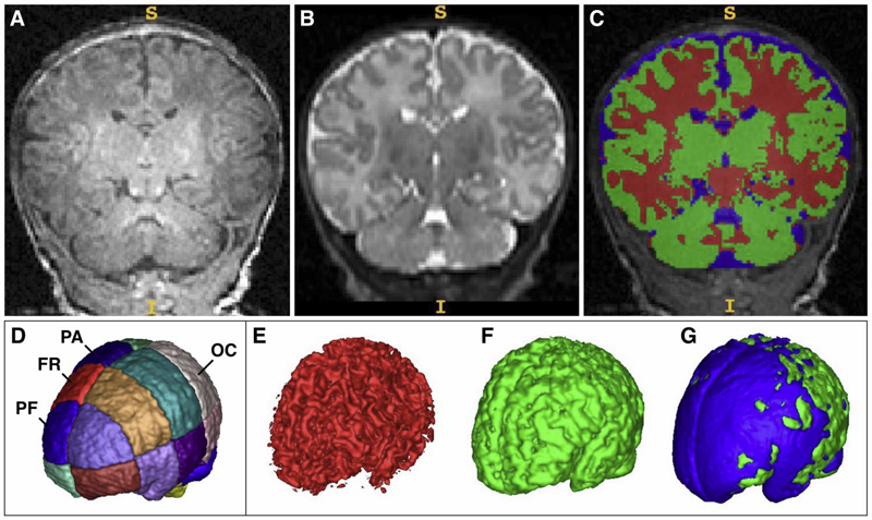
The brain undergoes rapid development during early childhood as a series of biophysical and chemical processes occur, which can be observed in magnetic resonance (MR) images as a change over time of white matter intensity relative to gray matter. Such a contrast change manifests in specific patterns in different imaging modalities, suggesting that brain maturation is encoded by appearance changes in multi-modal MRI. In this paper, we explore the patterns of early brain growth encoded by multi-modal contrast changes in a longitudinal study of children. For a given modality, contrast is measured by comparing histograms of intensity distributions between white and gray matter. Multivariate non-linear mixed effects (NLME) modeling provides subject-specific as well as population growth trajectories which accounts for contrast from multiple modalities. The multivariate NLME procedure and resulting non-linear contrast functions enable the study of maturation in various regions of interest. Our analysis of several brain regions in a study of 70 healthy children reveals a posterior to anterior pattern of timing of maturation in the major lobes of the cerebral cortex, with posterior regions maturing earlier than anterior regions. Furthermore, we find significant differences between maturation rates between males and females.
J. J. Wolff, M. R. Swanson, J. T. Elison, G. Gerig, J. R. Pruett, M. A. Styner, C. Vachet, K. N. Botteron, S. R. Dager, A. M. Estes, H. C. Hazlett, R. T. Schultz, M. D. Shen, L. Zwaigenbaum, J. Piven.
“Neural circuitry at age 6~months associated with later repetitive behavior and sensory responsiveness in autism,” In Molecular Autism, Vol. 8, No. 1, Springer Nature, March, 2017.
DOI: 10.1186/s13229-017-0126-z
Background
Restricted and repetitive behaviors are defining features of autism spectrum disorder (ASD). Under revised diagnostic criteria for ASD, this behavioral domain now includes atypical responses to sensory stimuli. To date, little is known about the neural circuitry underlying these features of ASD early in life.
Methods
Longitudinal diffusion tensor imaging data were collected from 217 infants at high familial risk for ASD. Forty-four of these infants were diagnosed with ASD at age 2. Targeted cortical, cerebellar, and striatal white matter pathways were defined and measured at ages 6, 12, and 24 months. Dependent variables included the Repetitive Behavior Scale-Revised and the Sensory Experiences Questionnaire.
Results
Among children diagnosed with ASD, repetitive behaviors and sensory response patterns were strongly correlated, even when accounting for developmental level or social impairment. Longitudinal analyses indicated that the genu and cerebellar pathways were significantly associated with both repetitive behaviors and sensory responsiveness but not social deficits. At age 6 months, fractional anisotropy in the genu significantly predicted repetitive behaviors and sensory responsiveness at age 2. Cerebellar pathways significantly predicted later sensory responsiveness. Exploratory analyses suggested a possible disordinal interaction based on diagnostic status for the association between fractional anisotropy and repetitive behavior.
Conclusions
Our findings suggest that restricted and repetitive behaviors contributing to a diagnosis of ASD at age 2 years are associated with structural properties of callosal and cerebellar white matter pathways measured during infancy and toddlerhood. We further identified that repetitive behaviors and unusual sensory response patterns co-occur and share common brain-behavior relationships. These results were strikingly specific given the absence of association between targeted pathways and social deficits.
2016
S. Elhabian, C. Vachet, J. Piven, M. Styner, G. Gerig.
“Compressive sensing based Q-space resampling for handling fast bulk motion in hardi acquisitions,” In 2016 IEEE 13th International Symposium on Biomedical Imaging (ISBI), IEEE, pp. 907--910. April, 2016.
DOI: 10.1109/isbi.2016.7493412
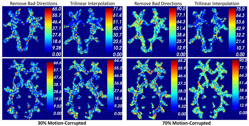
Diffusion-weighted (DW) MRI has become a widely adopted imaging modality to reveal the underlying brain connectivity. Long acquisition times and/or non-cooperative patients increase the chances of motion-related artifacts. Whereas slow bulk motion results in inter-gradient misalignment which can be handled via retrospective motion correction algorithms, fast bulk motion usually affects data during the application of a single diffusion gradient causing signal dropout artifacts. Common practices opt to discard gradients bearing signal attenuation due to the difficulty of their retrospective correction, with the disadvantage to lose full gradients for further processing. Nonetheless, such attenuation might only affect limited number of slices within a gradient volume. Q-space resampling has recently been proposed to recover corrupted slices while saving gradients for subsequent reconstruction. However, few corrupted gradients are implicitly assumed which might not hold in case of scanning unsedated infants or patients in pain. In this paper, we propose to adopt recent advances in compressive sensing based reconstruction of the diffusion orientation distribution functions (ODF) with under sampled measurements to resample corrupted slices. We make use of Simple Harmonic Oscillator based Reconstruction and Estimation (SHORE) basis functions which can analytically model ODF from arbitrary sampled signals. We demonstrate the impact of the proposed resampling strategy compared to state-of-art resampling and gradient exclusion on simulated intra-gradient motion as well as samples from real DWI data.
S. Kim, I.Lyu, V. Fonov, C. Vachet, H. Hazlett, R. Smith, J. Piven, S. Dager, R. Mckinstry, J. Pruett, A. Evans, D. Collins, K. Botteron, R. Schultz, G. Gerig, M. Styner.
“Development of Cortical Shape in the Human Brain from 6 to 24 Months of Age via a Novel Measure of Shape Complexity,” In NeuroImage, Vol. 135, Elsevier, pp. 163--176. July, 2016.
DOI: 10.1016/j.neuroimage.2016.04.053
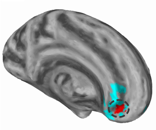
The quantification of local surface morphology in the human cortex is important for examining population differences as well as developmental changes in neurodegenerative or neurodevelopmental disorders. We propose a novel cortical shape measure, referred to as the 'shape complexity index' (SCI), that represents localized shape complexity as the difference between the observed distributions of local surface topology, as quantified by the shape index (SI) measure, to its best fitting simple topological model within a given neighborhood. We apply a relatively small, adaptive geodesic kernel to calculate the SCI. Due to the small size of the kernel, the proposed SCI measure captures fine differences of cortical shape. With this novel cortical feature, we aim to capture comparatively small local surface changes that capture a) the widening versus deepening of sulcal and gyral regions, as well as b) the emergence and development of secondary and tertiary sulci. Current cortical shape measures, such as the gyrification index (GI) or intrinsic curvature measures, investigate the cortical surface at a different scale and are less well suited to capture these particular cortical surface changes. In our experiments, the proposed SCI demonstrates higher complexity in the gyral/sulcal wall regions, lower complexity in wider gyral ridges and lowest complexity in wider sulcal fundus regions. In early postnatal brain development, our experiments show that SCI reveals a pattern of increased cortical shape complexity with age, as well as sexual dimorphisms in the insula, middle cingulate, parieto-occipital sulcal and Broca's regions. Overall, sex differences were greatest at 6months of age and were reduced at 24months, with the difference pattern switching from higher complexity in males at 6months to higher complexity in females at 24months. This is the first study of longitudinal, cortical complexity maturation and sex differences, in the early postnatal period from 6 to 24months of age with fine scale, cortical shape measures. These results provide information that complement previous studies of gyrification index in early brain development.
2015
C.C. Conlin, J.L. Zhang, F. Rousset, C. Vachet, Y. Zhao, K.A. Morton, K. Carlston, G. Gerig, V.S. Lee.
“Performance of an Efficient Image-registration Algorithm in Processing MR Renography Data,” In J Magnetic Resonance Imaging, July, 2015.
DOI: 10.1002/jmri.25000
PURPOSE:
To evaluate the performance of an edge-based registration technique in correcting for respiratory motion artifacts in magnetic resonance renographic (MRR) data and to examine the efficiency of a semiautomatic software package in processing renographic data from a cohort of clinical patients.
MATERIALS AND METHODS:
The developed software incorporates an image-registration algorithm based on the generalized Hough transform of edge maps. It was used to estimate glomerular filtration rate (GFR), renal plasma flow (RPF), and mean transit time (MTT) from 36 patients who underwent free-breathing MRR at 3T using saturation-recovery turbo-FLASH. The processing time required for each patient was recorded. Renal parameter estimates and model-fitting residues from the software were compared to those from a previously reported technique. Interreader variability in the software was quantified by the standard deviation of parameter estimates among three readers. GFR estimates from our software were also compared to a reference standard from nuclear medicine.
RESULTS:
The time taken to process one patient's data with the software averaged 12 ± 4 minutes. The applied image registration effectively reduced motion artifacts in dynamic images by providing renal tracer-retention curves with significantly smaller fitting residues (P < 0.01) than unregistered data or data registered by the previously reported technique. Interreader variability was less than 10% for all parameters. GFR estimates from the proposed method showed greater concordance with reference values (P < 0.05).
CONCLUSION:
These results suggest that the proposed software can process MRR data efficiently and accurately. Its incorporated registration technique based on the generalized Hough transform effectively reduces respiratory motion artifacts in free-breathing renographic acquisitions. J. Magn. Reson. Imaging 2015.
I. OguzI, J. Cates, M. Datar, B. Paniagua, T. Fletcher, C. Vachet, M. Styner, R. Whitaker.
“Entropy-based particle correspondence for shape populations,” In International Journal of Computer Assisted Radiology and Surgery, Springer, pp. 1-12. December, 2015.
Purpose
Statistical shape analysis of anatomical structures plays an important role in many medical image analysis applications such as understanding the structural changes in anatomy in various stages of growth or disease. Establishing accurate correspondence across object populations is essential for such statistical shape analysis studies.
Methods
In this paper, we present an entropy-based correspondence framework for computing point-based correspondence among populations of surfaces in a groupwise manner. This robust framework is parameterization-free and computationally efficient. We review the core principles of this method as well as various extensions to deal effectively with surfaces of complex geometry and application-driven correspondence metrics.
Results
We apply our method to synthetic and biological datasets to illustrate the concepts proposed and compare the performance of our framework to existing techniques.
Conclusions
Through the numerous extensions and variations presented here, we create a very flexible framework that can effectively handle objects of various topologies, multi-object complexes, open surfaces, and objects of complex geometry such as high-curvature regions or extremely thin features.
A. P. Salzwedel, K. M. Grewen, C. Vachet, G. Gerig, W. Lin,, W. Gao.
“Prenatal Drug Exposure Affects Neonatal Brain Functional Connectivity,” In The Journal of Neuroscience, Vol. 35, No. 14, pp. 5860-5869. April, 2015.
DOI: 10.1523/JNEUROSCI.4333-14.2015
M. R. Swanson, J. J. Wolff, J. T. Elison, H. Gu, H. C. Hazlett, K. Botteron, M. Styner, S. Paterson, G. Gerig, J. Constantino, S. Dager, A. Estes, C. Vachet, J. Piven.
“Splenium development and early spoken language in human infants,” In Developmental Science, Wiley Online Library, 2015.
ISSN: 1467-7687
DOI: 10.1111/desc.12360
The association between developmental trajectories of language-related white matter fiber pathways from 6 to 24 months of age and individual differences in language production at 24 months of age was investigated. The splenium of the corpus callosum, a fiber pathway projecting through the posterior hub of the default mode network to occipital visual areas, was examined as well as pathways implicated in language function in the mature brain, including the arcuate fasciculi, uncinate fasciculi, and inferior longitudinal fasciculi. The hypothesis that the development of neural circuitry supporting domain-general orienting skills would relate to later language performance was tested in a large sample of typically developing infants. The present study included 77 infants with diffusion weighted MRI scans at 6, 12 and 24 months and language assessment at 24 months. The rate of change in splenium development varied significantly as a function of language production, such that children with greater change in fractional anisotropy (FA) from 6 to 24 months produced more words at 24 months. Contrary to findings from older children and adults, significant associations between language production and FA in the arcuate, uncinate, or left inferior longitudinal fasciculi were not observed. The current study highlights the importance of tracing brain development trajectories from infancy to fully elucidate emerging brain–behavior associations while also emphasizing the role of the splenium as a key node in the structural network that supports the acquisition of spoken language.
J. J. Wolff, G. Gerig, J. D. Lewis, T. Soda, M. A. Styner, C. Vachet, K. N. Botteron, J. T. Elison, S. R. Dager, A. M. Estes, H. C. Hazlett, R. T. Schultz, L. Zwaigenbaum, J. Piven.
“Altered corpus callosum morphology associated with autism over the first 2 years of life,” In Brain, 2015.
DOI: 10.1093/brain/awv118

2014
S. Elhabian, Y. Gur, C. Vachet, J. Piven, M. Styner, I. Leppert, G.B. Pike, G. Gerig.
“A Preliminary Study on the Effect of Motion Correction On HARDI Reconstruction,” In Proceedings of the 2014 IEEE International Symposium on Biomedical Imaging (ISBI), pp. (accepted). 2014.
Keywords: Diffusion MRI, HARDI, motion correction, interpolation
S.Y. Elhabian, Y. Gur, C. Vachet, J. Piven, M.A. Styner, I.R. Leppert, B. Pike, G. Gerig.
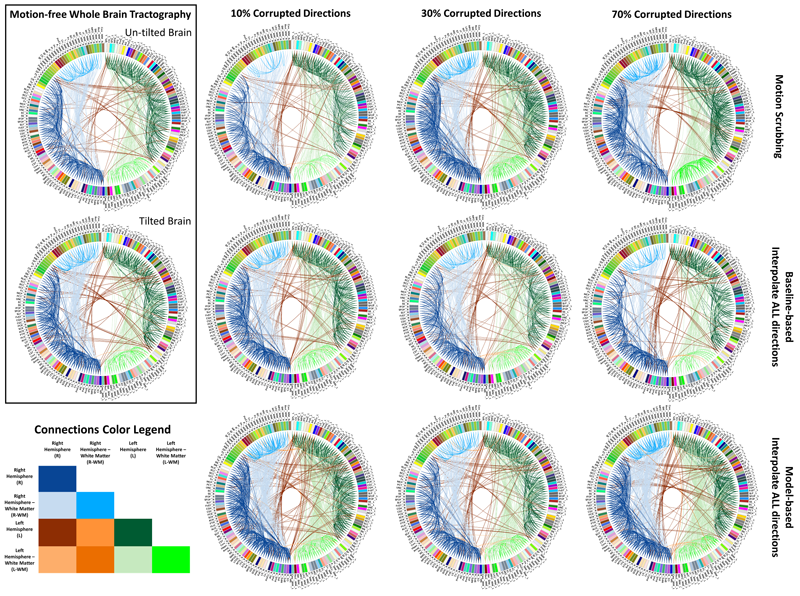
“Subject-Motion Correction in HARDI Acquisitions: Choices and Consequences,” In Frontiers in Neurology - Brain Imaging Methods, 2014.
DOI: 10.3389/fneur.2014.00240
K. Grewen, M. Burchinal, C. Vachet, S. Gouttard, J.H. Gilmore, W. Lin, J. Johns, M. Elam, G. Gerig.
“Prenatal cocaine effects on brain structure in early infancy,” In NeuroImage, Vol. 101, pp. 114--123. November, 2014.
DOI: 10.1016/j.neuroimage.2014.06.070
F. Rousset, C. Vachet, C. Conlin, M. Heilbrun, J.L. Zhang, V.S. Lee, G. Gerig.
“Semi-automated application for kidney motion correction and filtration analysis in MR renography,” In Proceeding of the 2014 Joint Annual Meeting ISMRM-ESMRMB, pp. (accepted). 2014.
Altered renal function commonly affects patients with cirrhosis, a consequence of chronic liver disease. From lowdose contrast material-enhanced magnetic resonance (MR) renography, we can estimate the Glomerular Filtration Rate (GFR), an important parameter to assess renal function. Two-dimensional MR images are acquired every 2 seconds for approximately 5 minutes during free breathing, which results in a dynamic series of 140 images representing kidney filtration over time. This specific acquisition presents dynamic contrast changes but is also challenged by organ motion due to breathing. Rather than use conventional image registration techniques, we opted for an alternative method based on object detection. We developed a novel analysis framework available under a stand-alone toolkit to efficiently register dynamic kidney series, manually select regions of interest, visualize the concentration curves for these ROIs, and fit them into a model to obtain GFR values. This open-source cross-platform application is written in C++, using the Insight Segmentation and Registration Toolkit (ITK) library, and QT4 as a graphical user interface.