High-Resolution Source Imaging from EEG
Collaborating Investigator: Dr. Don Tucker, Electrical Geodesics Inc. and University of OregonIn collaboration with Dr. Don Tucker and his colleagues at Electrical Geodesics Inc and the University of Oregon, the goal of this DBP is twofold: to improve our ability to reconstruct neuroelectric sources (source localization) from EEG measurements and to stimulate specific brain regions using electrodes attached on to the scalp transcranial direct current stimulation, tDCS).
For both research and clinical practice, EEG is a cost-effective tool to understand the macroscopic features of brain activity. Recent advances have significantly improved the spatial resolution of source estimates based on the EEG and offer the promise of precise spatio-temporal monitoring. Applying carefully controlled and precisely placed electric current by means of scalp electrodes can also stimulate cortical brain activity, which appears to alleviate symptoms for some neurological disorders. EEG technology, even advanced, high-resolution forms, is affordable even for small hospitals in remote locations and can be easily managed by technicians in the field.
Within the broad scope of research activity in the field of neural source imaging and stimulation, Dr. Tucker and CIBC have chosen a selected subset of projects with the goal of maximizing the impact of our collaborative research in terms of: (1) improving near-term translational uses of EEG-based source localization; (2) determining the spatial resolution ultimately possible with EEG; (3) improving the capabilities of our other neuroscience collaborators; and (4) being able to stimulate specific brain regions in a predefined manner and testing these results to address specific clinical problems.
The algorithms and tools developed in this project will be of immediate use to EGI and Dr. Tucker, and we expect to be able to achieve immediate and broad-based dissemination through their base of more than 400 users of their technology as well as CIBC's software dissemination mechanisms. In addition, our ability to collaboratively assert the true potential resolution of EEG in source imaging and noninvasive brain stimulation could make a very positive contribution to the adoption of these relatively low-cost technologies in many research and clinical laboratories.
Brain Connectivity Maps
This goal of this project is to develop tools for reconstruction of brain connectivity maps, and compare connectivity brain graphs using a novel metric. As was shown by Hammond etal1, structural brain connectivity networks can help improve accuracy in EEG source localization. The so-called "connectome matrices" that capture this connectivity can be used as priors, in order to stabilize the source localization solution. The CIBC role in this project is to reconstruct of brain connectivity networks using a previously developed method for extracting fiber orientations based on a tensor decomposition approach2. We are also developing a metric to compare brain networks in order to quantify differences in brain connectivity networks reconstructed form longitudinal measurements from the same patient.Fiber Tractography Visualization Tool
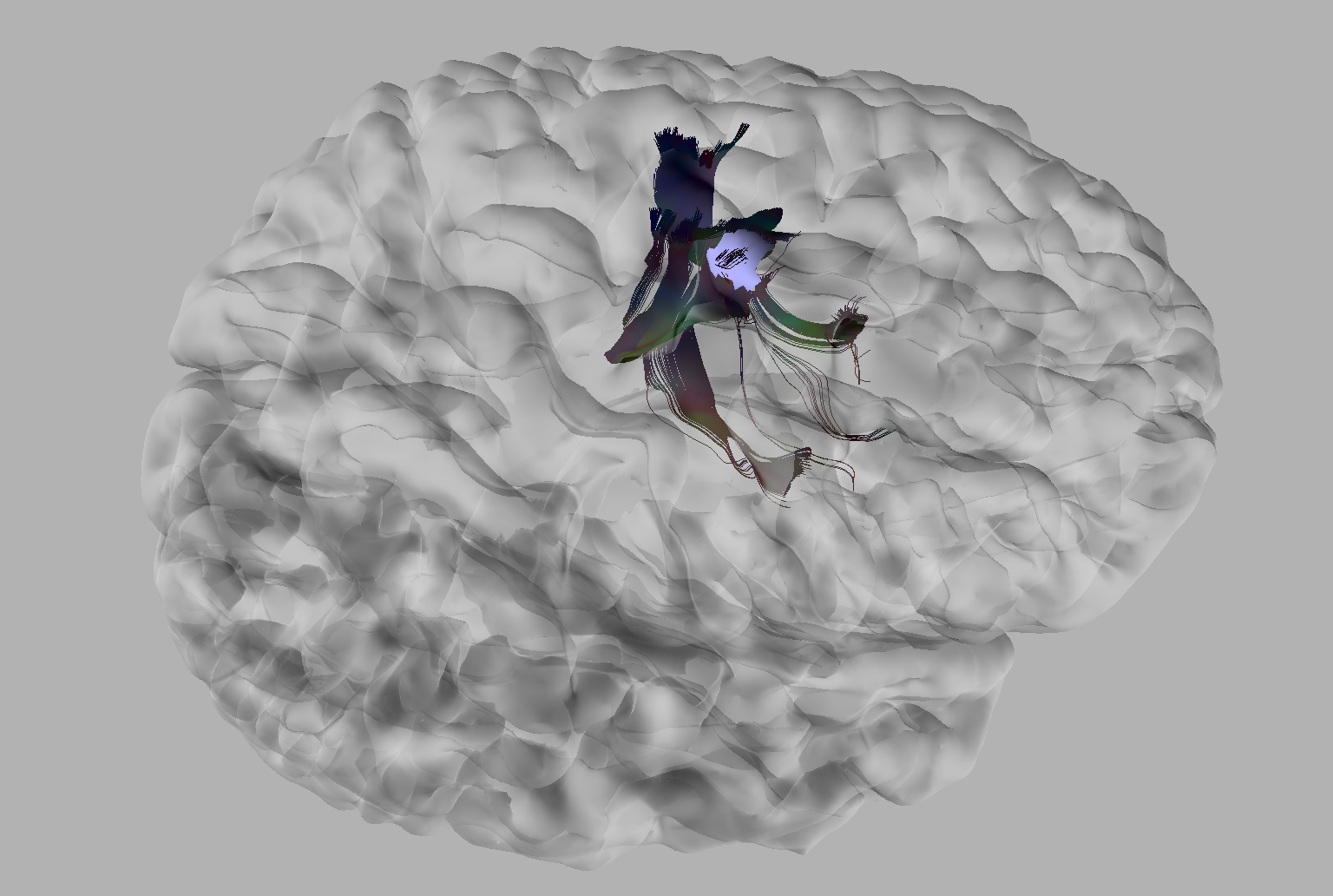
Dr. Tucker's laboratory at EGI provided a complete description of the data used to create the connectivity matrix. After providing EGI scientists with a tool capable of fusing these data into a single, coherent visualization, we adapted our existing multimodal data visualization tool to allow the interaction with larger datasets. This new suite of tools is capable of visualizing arbitrarily large white matter fiber tractography datasets through a streaming renderer. Additional improvements support pruning of these data by limiting the overall length of tracts shown. To allow scientists to more quickly and thoroughly interrogate these data, user-driven filtering has also been implemented. Figure shows how users may select regions of the cortex to highlight fiber bundles innervating those locations.Transcranial Direct Current Stimulation (tDCS)
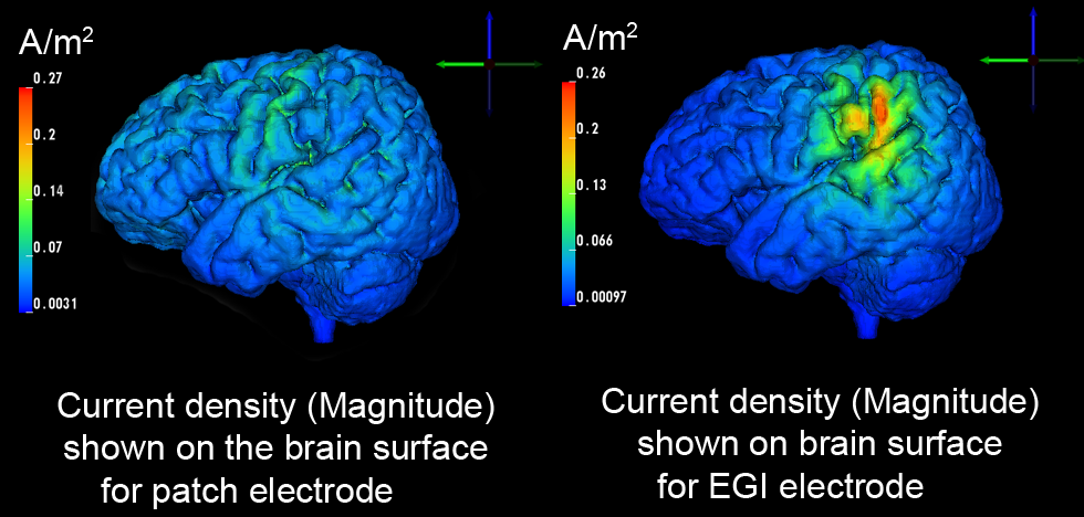
In certain clinical situations (treatment of mood disorders, enhance learning, etc, it is desirable to deliver low amplitude electrical stimulation currents to a particular brain region in a specific direction, while limiting current delivery to the rest of the brain. In this DBP we have built a system to simulate and study this translationally important technology. The driving hypothesis for the project is that using high-density (HD) electrode arrays rather than two conventional patch electrodes increases the ability to control the current patterns in the brain. With recent modifications of CIBC software (Seg3D, BioMesh3D, and SCIRun), we can simulate and study this capability using a subject-specific model of the head as an electrical conductor model3. In this way, we have confirmed that HD electrode arrays offer a more flexible means by which to stimulate particular brain regions compared to commonly used patch electrodes, while also being able to also replicate results achieved with those patch electrodes. Our results show that HD electrode arrays are capable of mimicking both spatially broad (Figure, left) and also focal, brain stimulations (Figure , right) while guaranteeing the safety of the subject. With the potential of this approach established, our emphasis has now switched to determining the best electrode injection patter to maximize the stimulation in a desired region.To meet this need, we have developed an optimization procedure that achieves the best possible delivery of current in a prespecified direction to a prespecified region of interest (ROI), while imposing safety constraints such as limiting the current delivered to the rest of the brain or at the skin surface. Early results suggest that we are capable of stimulating the ROI for an arbitrary direction. Additionally, it is important to show that the solution appears to be focal. Figure shows a histogram of the current intensity achieved in different anatomical regions throughout the brain for an optimized stimulation protocol. The current density in the ROI appears to be higher on average compared to the rest of the brain, although some regions also exhibit even higher densities . The level of specificity improves for ROIs close to the head surface, a well known challenge of external stimulation of the brain. Lastly, we are investigating the effect of tissue anisotropy in complex volume conductor models3 on the current optimization. Our initial findings suggest that skull and white matter anisotropy change the solution significantly depending on the desired stimulation direction within the ROI. In the coming year, we will focus on the comparison of traditional stimulation using patch electrodes with novel stimulation paradigms using HD electrode arrays and further optimization of current injection pattern for deep regions within the brain.
References:
1. D.K. Hammond, B. Scherrer, and A. Malony. Incorporating anatomical connectivity into EEG source estimation via sparse approximation with cortical graph wavelets. In IEEE Conference on Acoustics Signals and Signal Processing, 2012.2. F. Jiao, Y. Gur, C.R. Johnson, and S. Joshi. Detection of crossing white matter fibers with highorder tensors and rank-k decompositions. In Proceedings of the International Conference on Information Processing in Medical Imaging (IPMI 2011), volume 6801 of Lecture Notes in Computer Science (LNCS), pages 538–549, 2011.
3. M. Dannhauer, D.H. Brooks, D. Tucker, and R.S. MacLeod. A pipeline for the simulation of transcranial direct current stimulation for realistic human head models using SCIRun/BioMesh3D. In Proceedings of the 2012 IEEE Int. Conf. Engineering and Biology Society (EMBC), pages 5486–5489, 2012.


