2024
J.A. Bergquist, B. Zenger, J. Brundage, R.S. MacLeod, T.J. Bunch, R. Shah, X. Ye, A. Lyons, M. Torre, R. Ranjan, T. Tasdizen, B.A. Steinberg.
“Performance of Off-the-Shelf Machine Learning Architectures and Biases in Low Left Ventricular Ejection Fraction Detection,” In Heart Rhythm O2, Vol. 5, No. 9, pp. 644 - 654. 2024.
J.A. Bergquist, D. Dade, B. Zenger, R.S. MacLeod, X. Ye, R. Ranjan, T. Tasdizen, B.A. Steinberg.
“Machine Learning Prediction of Blood Potassium at Different Time Cutoffs,” In Computing in Cardiology 2024, 2024.
Because serum potassium and ECG morphology changes exhibit a well-understood connection, and the timeline of ECG changes can be relatively quick, there is motivation to explore the sensitivity of ML based prediction of serum potassium using 12 lead ECG data with respect to the time between the ECG and potassium readings.
We trained a convolutional neural network to classify abnormal (serum potassium above 5 mEq/L) vs normal (serum potassium between 4 and 5 mEq/L) from the ECG alone. We compared training with ECGs and potassium measurements filtered to be within 1 hour, 30 minutes, and 15 minutes of each other. We explored scenarios that both leveraged all available data at each time cutoff as well as restricted data to match training set sizes across the time cutoffs. For each case, we trained five separate instances of our neural network to account for variability.
The 1 hour cutoff with all data resulted in an average area under the receiver operator curve (AUC) of 0.850 and a weighted accuracy of 76.3%, 15 minutes resulted in 0.814, 72.5%, and 30 minutes. Truncating the training sets to the same size as the 15 minute cutoff results in comparable average accuracy and AUC for all. Our future studies will continue to explore the performance of ML potassium predictions through investigations of failure cases, identification of biases, and explainability analyses.
2013
C. Jones, M. Seyedhosseini, M. Ellisman, T. Tasdizen.
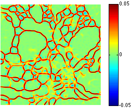
“Neuron Segmentation in Electron Microscopy Images Using Partial Differential Equations,” In Proceedings of 2013 IEEE 10th International Symposium on Biomedical Imaging (ISBI), pp. 1457--1460. April, 2013.
DOI: 10.1109/ISBI.2013.6556809

2010
J.R. Anderson, B.C. Grimm, S. Mohammed, B.W. Jones, T. Tasdizen, J. Spaltenstein, P. Koshevoy, R.T. Whitaker, R.E. Marc.
“The Viking Viewer: Scalable Multiuser Annotation and Summarization of Large Volume Datasets,” In Journal of Microscopy, Vol. 241, No. 1, pp. 13--28. 2010.
DOI: 10.1111/j.1365-2818.2010.03402.x
S. Gerber, T. Tasdizen, P.T. Fletcher, S. Joshi, R.T. Whitaker, the Alzheimers Disease Neuroimaging Initiative (ADNI).
“Manifold modeling for brain population analysis,” In Medical Image Analysis, Special Issue on the 12th International Conference on Medical Image Computing and Computer Assisted Intervention (MICCAI) 2009, Vol. 14, No. 5, Note: Awarded MICCAI 2010, Best of the Journal Issue Award, pp. 643--653. 2010.
ISSN: 1361-8415
DOI: 10.1016/j.media.2010.05.008
PubMed ID: 20579930
S.K. Iyer, E. DiBella, T. Tasdizen.
“Edge enhanced spatio-temporal constrained reconstruction of undersampled dynamic contrast enhanced radial MRI,” In IEEE International Symposium on Biomedical Imaging (ISBI): From Nano to Macro, pp. 704--707. 2010.
DOI: 10.1109/ISBI.2010.5490077
2007
G. Adluru, S.P. Awate, T. Tasdizen, R.T. Whitaker, E.V.R. DiBella.
“Temporally Constrained Reconstruction of Dynamic Cardiac Perfusion MRI,” In Magnetic Resonance in Medicine, Vol. 57, pp. 1027--1036. 2007.
2006
S.P. Awate, T. Tasdizen, R.T. Whitaker.
“Unsupervised Texture Segmentation with Nonparametric Neighborhood Statistics,” SCI Institute Technical Report, No. UUSCI-2006-011, University of Utah, 2006.
E. Jurrus, T. Tasdizen, P. Koshevoy, P.T. Fletcher, M. Hardy, C-B. Chien, W. Denk, R.T. Whitaker.
“Axon Tracking in Serial Block-Face Scanning Electron Microscopy,” In Workshop on Microscopic Image Analysis with Applications in Biology, MICCAI, October, 2006.
2005
T. Tasdizen, S.P. Awate, R.T. Whitaker, N. Foster.
“MRI Tissue Classification with Neighborhood Statistics: A Nonparametric, Entropy-Minimizing Approach,” In Proceedings of The 8th International Conference on Medical Image Computing and Computer Assisted Intervention (MICCAI), pp. 517--525. 2005.
PubMed ID: 16685999
2004
T. Tasdizen, D.M. Weinstein, J.N. Lee.
“Automatic Tissue Classification for the Human Head from Multispectral MRI,” SCI Institute Technical Report, No. UUSCI-2004-001, University of Utah, March, 2004.
T. Tasdizen, R.T. Whitaker.
“Higher-order nonlinear priors for surface reconstruction,” In IEEE Trans. Pattern Anal. & Mach. Intel., Vol. 26, No. 7, pp. 878--891. July, 2004.