 Sponsored by the Burton Foundation
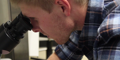
This summer, two Salt Lake area high school students from Copper Hills High School came to the University of Utah to participate in a hands-on research experience. The students learned how image analysis tools help biomechanics researchers understand the effects of structural features of musculoskeletal tissues (e.g. tendons, ligaments, and articular cartilage) on the functional behavior of these tissues.Many musculoskeletal tissue injuries and diseases exhibit altered macroscopic and microscopic tissue structure. The Musculoskeletal Research Laboratories, a research center of the Scientific Computing and Imaging Institute, uses engineered tissue materials to study the effect of these structural changes on tissue behavior. Researchers use many image acquisition techniques to characterize the structure of native and engineered tissues, including optical microscopy, x-ray computed tomography (CT), and electron microscopy. Image analysis tools allow efficient detection and quantification of structural features from these images.
The students in this summer's research intern program used state-of-the-art software tools to analyze the size and density of pores (empty space) in engineered tissue materials. For example, the students used ImageJ (http://imagej.nih.gov/ij/) to reconstruct large images from series of individual microscopy images. These techniques allow researchers to perform high-magnification imaging of large areas (e.g. entire tissues and organs) that are much bigger than a single field of view. Using Seg3D (http://sci.utah.edu/software/seg3d.html) the students generated 3D volumes from the reconstructed microscopy images. The students then experimented with a variety of image processing methods to isolate the structural features in the 3D image volumes, performing statistical analysis on their processed image volumes to compare the different groups of engineered tissues. Throughout this process, the students had to expand their knowledge base, learning about methods of image reconstruction, digital image filters, and computer programming.
In this internship, the students applied these image processing and analysis techniques to investigate engineered tissue materials, but this is not the limit of the skills they learned this summer. Digital image analysis has broad application throughout biomedicine clinical practice and research (for example, studying heart, brain, and lung structure and function) as well as outside of medicine (for example, detecting changes in forestation from satellite images and tracking atmospheric weather patterns). Both of our summer internship students gained valuable experience with the tools of biomedical engineers and researchers as they explore their options beyond high school and developed skills applicable across a wide range of scientific areas.