Software Tools for Image Based Modeling, Simulation, and Visualization
August 31st, 2011at EMBS 2011
Boston, MA
Summary
The goal of this tutorial is to introduce participants to a suite of software tools for image-based modeling, simulation, and visualization developed by the NIH/NCRR Center for Integrative Biomedical Computing (CIBC). This portable and flexible collection of interactive tools was designed in particular to support the development of subject specific, image based geometric models for simulation of bioelectric fields. The tools, individually or as a suite, have much broader utility and have supported research and clinical projects all over the world.
The tools in the suite include:
- Seg3D, for general purpose user-guided image segmentation;
- ImageVis3D, for visualization of large scale data;
- map3d, for visualization of surface based maps from multichannel time signals;
- BioMesh3D, a set of utilities for creating surface and volume meshes from segmented image data;
- SCIRun, a comprehensive problem solving environment that integrates many of the capabilities of an entire image based modeling pipeline.
The tutorial will be a mix of didactic presentations on the components of image based modeling, simulation, and visualization; hands on practice with the software; and cases studies of real world applications. We will provide participants with the software and test data sets and encourage participants to bring their laptop computers, and, if relevant, their own data. CIBC members and developers will be on hand to help participants learn the programs, port their data, and generate useful results. We especially encourage participation by students, post docs, and technical users and software developers. Binary distribution installers (recommended) will be available for Apple OS X and Windows operating systems.
Presentations
1.1 Introduction (Rob MacLeod, Dana Brooks)
1.2 Case Study I: Image based analysis of patients with atrial fibrillation (Rob MacLeod)
1.3 Demo I: Seg3D Demo and tutorial (Jess Tate)
1.4 Lab I: Segmentation with Seg3D
1.5 Case Study II: Virtual Comparison of Deep Brain Stimulation Parameters (Tom Fogal)
1.6 Demo II: ImageVis3D/map3d demo and tutorial (Tom Fogal & Burak Erem)
ImageVis3D Software & Data
ImageVis3D Tutorial (pdf)
Getting Data Into ImageVis3D (pdf)
ImageVis3D Documentation (pdf)
map3d Software
1.7 Lab II: Visualization with ImageVis3D and map3D
1.8 Case Study III: Modeling of cardiac ischemia and arrhythmia (Rob MacLeod)
1.9 Demo III: BioMesh3D demo and tutorial (Derrell Swenson, Josh Levine)
SCIRun BioMesh3D Software & Data
1.10 Lab III: Mesh generation with BioMesh3D
1.11 Case Study IV: Simulation of defibrillation (Jess Tate)
1.12 Demo IV: SCIRun demo and tutorial (Jess Tate)
1.13 Lab IV: Simulation with SCIRun
CIBC Software Requirements
SCIRun
Graphics cards must support OpenGL 2.0 or greater.Windows: XP or better
OS X: 10.5, 10.6 (10.7 support is in progress)
Linux: SCIRun must be compiled from source and requires the following:
GCC 4 compiler
CMake 2.6 or greater
NVIDIA card and drivers
Installing platform-specific packages may be required.
SCIRun is known to compile on OpenSuSE 10 and 11, Ubuntu 9 and 10, and RHEL 5 (also CentOS and Fedora).
Seg3d
OSX minimum requirement: Mac OS X 10.5 or 10.6 (10.7 should work as well)Windows minimum requirement: Windows XP, Windows Vista, or Windows 7, Strongly recommended 64bit operating system
Linux minimum requirement: Only for advanced users: need to build from source (build can take up to an hour), but should work on any Linux flavor
CPU: minimum Core Duo or higher, recommended i5 or i7
Memory: minimum 4Gb, recommended 8Gb or more
Graphics Driver: minimum OpenGL 2.0 or higher (Not available on older Intel embedded graphics cards)
Graphics memory: 128, recommended 256Mb or more
BioMesh3D
Graphics cards must support OpenGL 2.0 or greater.Python is required to run the BioMesh3D pipeline.
Multicore processor is strongly recommended.
Windows: Vista or 7
OS X: 10.5, 10.6
Linux: BioMesh3D must be compiled from source and requires the following:
GCC 4 compiler
CMake 2.6 or greater
NVIDIA card and drivers
Installing platform-specific packages may be required.
BioMesh3D comes packaged with SCIRun, which is known to compile on:
- OpenSuSE 10 and 11
- Ubuntu 9 and 10
- RHEL 5
- CentOS
- Fedora
ImageVis3d
OS X: 10.6 or 10.7Windows: Windows 7 or Windows Vista
Linux: Debian, Ubuntu, or Red Hat, with NVIDIA card and proprietary drivers.
Hardware, GPU: NVIDIA GeForce 9 series or better (i.e. GeForce 300M or later), or ATI/AMD Radeon HD 2400 series or better
map3d
Graphics cards must support OpenGL 2.0 or better.Required: 1.5 GHz CPU, 1GB RAM, 10GB free disk space, 64MB video RAM
Recommended: Dual-core 2 GHz CPU, 2GB RAM, 10GB free disk space, 128MB video RAM
Operating systems: Windows XP, Vista, 7, Mac OSX 10.5 or greater
map3d can be built on many linux distributions (will require a C++ compiler and Qt SDK).
Extra Minisymposium at EMBS
Imaging and Computation in Clinical Electrocardiology
Thursday, September 1st from 5:15pm - 6:45pmMarriott 3rd floor, Berkeley
Open to all registered conference attendees
| Organizers: | Robert MacLeod, PhD - University of Utah Dana Brooks, PhD - Northeastern University |
| Speakers: | Ravi Ranjan, MD PhD - University of Utah Petr Stovicek, MD PhD - Charles University Hospital John Triedman, MD - Harvard Medical School |
Investigators have been conducting research on the use of imaging and computation in electrocardiology for many years. However until fairly recently the impact of advances in this area on clinical practice has been minimal except in a few specialized areas. In contrast, recent research progress, combined with improvements in imaging and computational technology itself, have begun to result in many exciting possibilities for clinical application of research results. In particular the combination of imaging and computation is enabling dramatic expansion of the concept of "personalized medicine" beyond the world of genomics by enabling patient-specific modeling and simulation. This minisymposium will focus on both progress and challenges in three distinct clinical applications of imaging and computational research, from the points of view of three presenters who are each clinicians who are active in both clinical practice and research.
Presenter bios:
Ravi Ranjan, MD, PhD, is Assistant Professor of Medicine, University of Utah. He is a clinical cardiac electrophysiologist with a keen interest in understanding arrhythmia mechanisms and developing new therapies. His PhD research was on understanding ion channel physiology and cardiac excitation and propagation using experimental studies and mathematical modeling. He continued work on ion channel structure and function during his medical studies. Recent work focused on the importance of gaps in ablation lesion sets as a cause of poor outcome of ablation procedures. Currently, he is working on better understanding of atrial fibrillation in an animal model using MRI.
Petr Stovicek, MD, PhD, has been engaged in ECG research since 1986, He has worked on simultaneous cardiac polygraphy and body surface potential mapping and has published on pathologies, including comparative case-control studies and invasive measurements in patient with coronary artery disease and arrhythmias. After training at Dalhousie University and QEII Health Sciences Centre in Halifax, Canada, he opened a cardiac electrophysiology laboratory for Charles University Hospital (Department of Medicine II) in Prague, Czech Republic, where he introduced routine catheter ablations of arrhythmias. More recently he has been advancing clinical use of biophysical computer modellng and imaging (ECG lead transformation, inverse electrocardiography).
John Triedman, MD, is Professor of Pediatrics at Harvard Medical School and a senior cardiologist at Children's Hospital Boston, specializing in interventional cardiac electrophysiology. His research topics have included three-dimensional arrhythmia mapping, the pathogenesis of arrhythmias in congenital heart disease, the application of nonfluoroscopic navigation to catheter ablation, and modeling of defibrillating electrical fields in the human thorax.
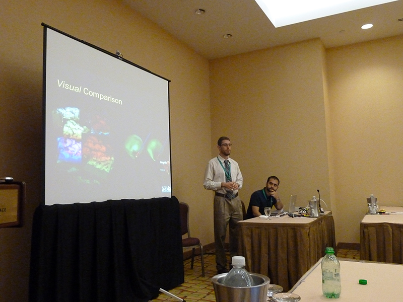
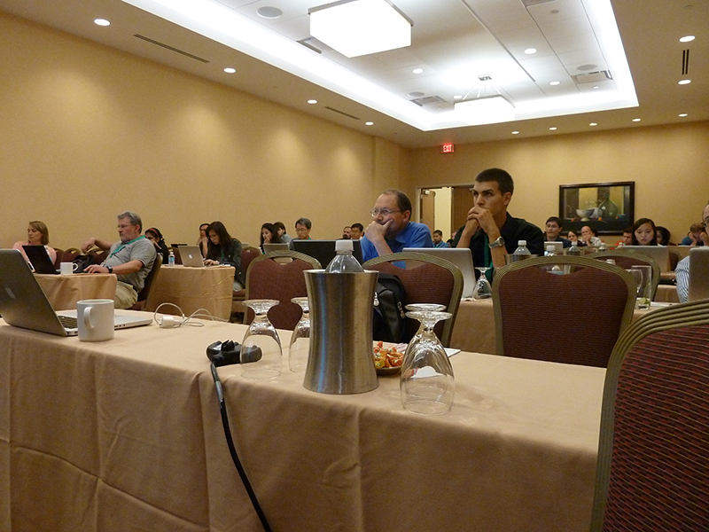
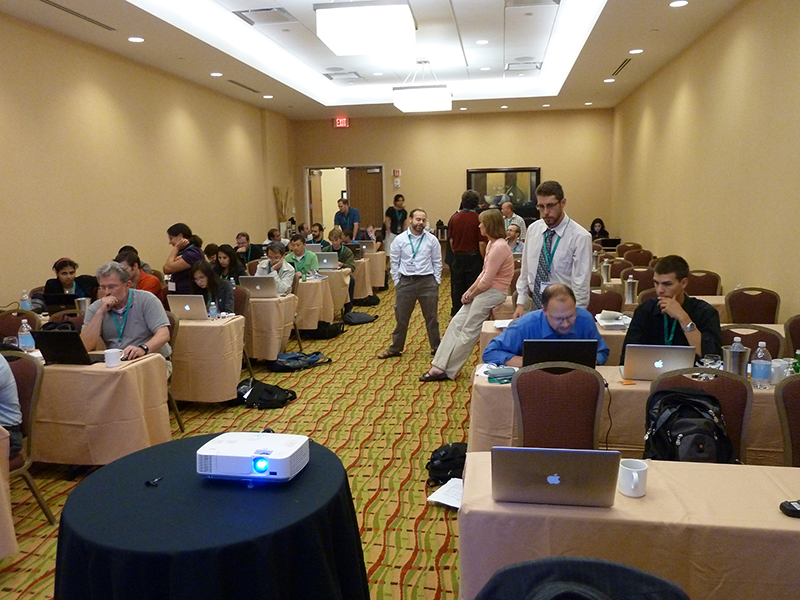
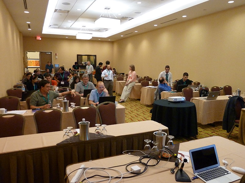
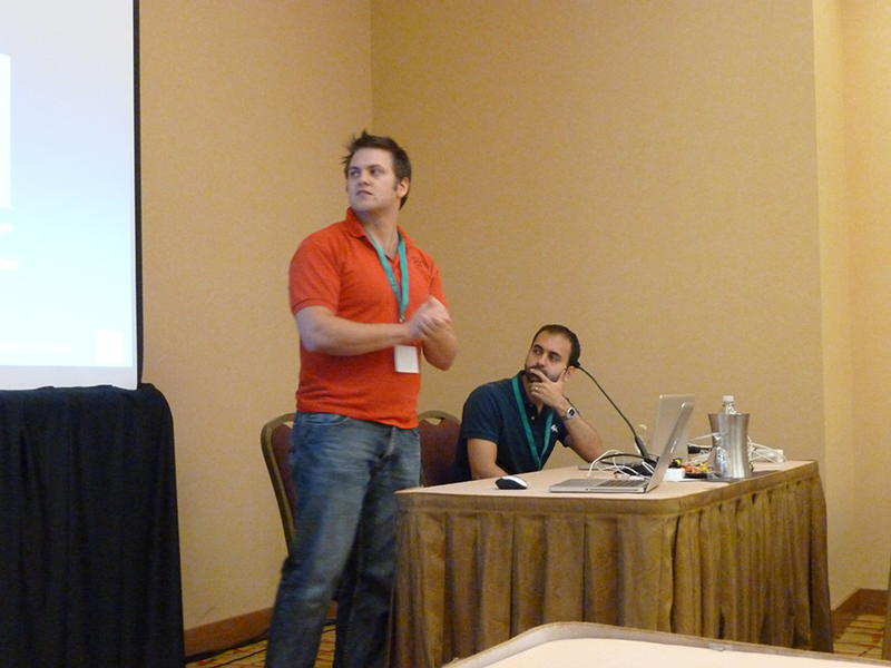
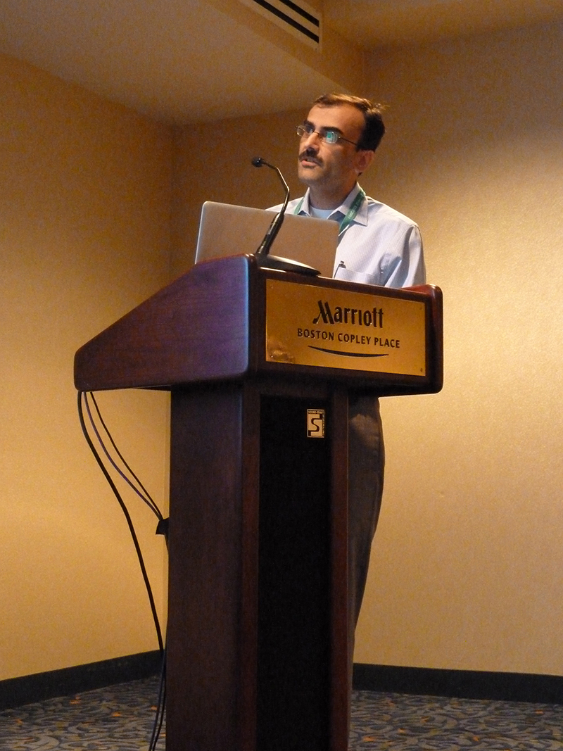
Photos
Thanks to all those who attended. We hope to see you back at our next workshop.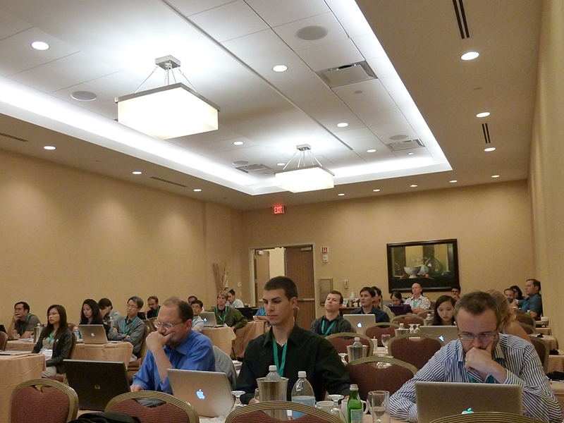 |
 |
 |
 |
 |
 |
 |
 |
 |
 |