


SCI Publications
2015


C. Nobre, A. Lex.
“OceanPaths: Visualizing Multivariate Oceanography Data,” In Eurographics Conference on Visualization (EuroVis) - Short Papers, Edited by E. Bertini, J. Kennedy, E. Puppo, The Eurographics Association, 2015.
DOI: 10.2312/eurovisshort.20151124
Geographical datasets are ubiquitous in oceanography. While map-based visualizations are useful for many different domains, they can suffer from cluttering and overplotting issues when used for multivariate data sets. As a result, spatial data exploration in oceanography has often been restricted to multiple maps showing various depths or time intervals. This lack of interactive exploration often hinders efforts to expose correlations between properties of oceanographic features, specifically currents. OceanPaths provides powerful interaction and exploration methods for spatial, multivariate oceanography datasets to remedy these situations. Fundamentally, our method allows users to define pathways, typically following currents, along which the variation of the high-dimensional data can be plotted efficiently. We present a case study conducted by domain experts to underscore the usefulness of OceanPaths in uncovering trends and correlations in oceanographic data sets.
I. OguzI, J. Cates, M. Datar, B. Paniagua, T. Fletcher, C. Vachet, M. Styner, R. Whitaker.
“Entropy-based particle correspondence for shape populations,” In International Journal of Computer Assisted Radiology and Surgery, Springer, pp. 1-12. December, 2015.
Purpose
Statistical shape analysis of anatomical structures plays an important role in many medical image analysis applications such as understanding the structural changes in anatomy in various stages of growth or disease. Establishing accurate correspondence across object populations is essential for such statistical shape analysis studies.
Methods
In this paper, we present an entropy-based correspondence framework for computing point-based correspondence among populations of surfaces in a groupwise manner. This robust framework is parameterization-free and computationally efficient. We review the core principles of this method as well as various extensions to deal effectively with surfaces of complex geometry and application-driven correspondence metrics.
Results
We apply our method to synthetic and biological datasets to illustrate the concepts proposed and compare the performance of our framework to existing techniques.
Conclusions
Through the numerous extensions and variations presented here, we create a very flexible framework that can effectively handle objects of various topologies, multi-object complexes, open surfaces, and objects of complex geometry such as high-curvature regions or extremely thin features.
B.R. Parmar, T.R. Jarrett, E.G. Kholmovski, N. Hu, D. Parker, R.S. MacLeod, N.F. Marrouche, R. Ranjan.
“Poor scar formation after ablation is associated with atrial fibrillation recurrence,” In Journal of Interventional Cardiac Electrophysiology, Vol. 44, No. 3, pp. 247-256. December, 2015.
Purpose
Patients routinely undergo ablation for atrial fibrillation (AF) but the recurrence rate remains high. We explored in this study whether poor scar formation as seen on late-gadolinium enhancement magnetic resonance imaging (LGE-MRI) correlates with AF recurrence following ablation.
Methods
We retrospectively identified 94 consecutive patients who underwent their initial ablation for AF at our institution and had pre-procedural magnetic resonance angiography (MRA) merged with left atrial (LA) anatomy in an electroanatomic mapping (EAM) system, ablated areas marked intraprocedurally in EAM, 3-month post-ablation LGE-MRI for assessment of scar, and minimum of 3-months of clinical follow-up. Ablated area was quantified retrospectively in EAM and scarred area was quantified in the 3-month post-ablation LGE-MRI.
Results
With the mean follow-up of 336 days, 26 out of 94 patients had AF recurrence. Age, hypertension, and heart failure were not associated with AF recurrence, but LA size and difference between EAM ablated area and LGE-MRI scar area was associated with higher AF recurrence. For each percent higher difference between EAM ablated area and LGE-MRI scar area, there was a 7–9 % higher AF recurrence (p values 0.001–0.003) depending on the multivariate analysis.
Conclusions
In AF ablation, poor scar formation as seen on LGE-MRI was associated with AF recurrence. Improved mapping and ablation techniques are necessary to achieve the desired LA scar and reduce AF recurrence.
B. Peterson, N. Xiao, J. Holmen, S. Chaganti, A. Pakki, J. Schmidt, D. Sunderland, A. Humphrey, M. Berzins.
“Developing Uintah’s Runtime System For Forthcoming Architectures,” Subtitled “Refereed paper presented at the RESPA 15 Workshop at SuperComputing 2015 Austin Texas,” SCI Institute, 2015.
B. Peterson, H. K. Dasari, A. Humphrey, J.C. Sutherland, T. Saad, M. Berzins.
“Reducing overhead in the Uintah framework to support short-lived tasks on GPU-heterogeneous architectures,” In Proceedings of the 5th International Workshop on Domain-Specific Languages and High-Level Frameworks for High Performance Computing (WOLFHPC'15), ACM, pp. 4:1-4:8. 2015.
DOI: 10.1145/2830018.2830023
J. M. Phillips, Bei Wang, Y. Zheng.
“Geometric Inference on Kernel Density Estimates,” In CoRR, Vol. abs/1307.7760, 2015.
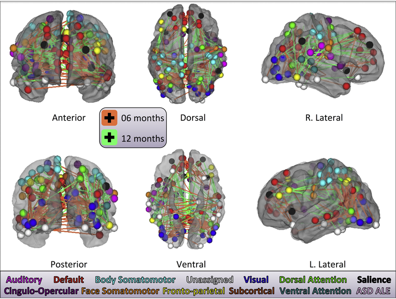
J.R. Pruett Jr., S. Kandala, S. Hoertel, A.Z. Snyder, J.T. Elison, T. Nishino, E. Feczko, N.U.F. Dosenbach, B. Nardos, J.D. Power, B. Adeyemo, K.N. Botteron, R.C. McKinstry, A.C. Evans, H.C. Hazlett, S.R. Dager, S. Paterson, R.T. Schultz, D.L. Collins, V.S. Fonov, M. Styner, G. Gerig, S. Das, P. Kostopoulos, J.N. Constantino, A.M. Estes, The IBIS Network, S.E. Petersen, B.L. Schlaggar, J. Piven.
“Accurate age classification of 6 and 12 month-old infants based on resting-state functional connectivity magnetic resonance imaging data,” In Developmental Cognitive Neuroscience, Vol. 12, pp. 123--133. April, 2015.
DOI: 10.1016/j.dcn.2015.01.003
S. Pujol, W. Wells, C. Pierpaoli, C. Brun, J. Gee, G. Cheng, B. Vemuri, O. Commowick, S. Prima, A. Stamm, M. Goubran, A. Khan, T. Peters, P. Neher, K. H. Maier-Hein, Y. Shi, A. Tristan-Vega, G. Veni, R. Whitaker, M. Styner, C.F. Westin, S. Gouttard, I. Norton, L. Chauvin, H. Mamata, G. Gerig, A. Nabavi, A. Golby,, R. Kikinis.
“The DTI Challenge: Toward Standardized Evaluation of Diffusion Tensor Imaging Tractography for Neurosurgery,” In Journal of Neuroimaging, Wiley, August, 2015.
DOI: 10.1111/jon.12283

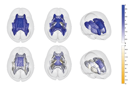
Diffusion tensor imaging (DTI) tractography reconstruction of white matter pathways can help guide brain tumor resection. However, DTI tracts are complex mathematical objects and the validity of tractography-derived information in clinical settings has yet to be fully established. To address this issue, we initiated the DTI Challenge, an international working group of clinicians and scientists whose goal was to provide standardized evaluation of tractography methods for neurosurgery. The purpose of this empirical study was to evaluate different tractography techniques in the first DTI Challenge workshop.
METHODS
Eight international teams from leading institutions reconstructed the pyramidal tract in four neurosurgical cases presenting with a glioma near the motor cortex. Tractography methods included deterministic, probabilistic, filtered, and global approaches. Standardized evaluation of the tracts consisted in the qualitative review of the pyramidal pathways by a panel of neurosurgeons and DTI experts and the quantitative evaluation of the degree of agreement among methods.
RESULTS
The evaluation of tractography reconstructions showed a great interalgorithm variability. Although most methods found projections of the pyramidal tract from the medial portion of the motor strip, only a few algorithms could trace the lateral projections from the hand, face, and tongue area. In addition, the structure of disagreement among methods was similar across hemispheres despite the anatomical distortions caused by pathological tissues.
CONCLUSIONS
The DTI Challenge provides a benchmark for the standardized evaluation of tractography methods on neurosurgical data. This study suggests that there are still limitations to the clinical use of tractography for neurosurgical decision making.
M. Raj, M. Mirzargar, R. Kirby, R. Whitaker, J. Preston.
“Evaluating Alignment of Shapes by Ensemble Visualization,” In IEEE Computer Graphics and Applications, IEEE, 2015.
The visualization of variability in 3D shapes or surfaces, which is a type of ensemble uncertainty visualization for volume data, provides a means of understanding the underlying distribution for a collection or ensemble of surfaces. While ensemble visualization for surfaces is already described in the literature, we conduct an expert-based evaluation in a particular medical imaging application: the construction of atlases or templates from a population of images. In this work, we extend contour boxplots to 3D, allowing us to evaluate it against an enumeration-style visualization of the ensemble members and also other conventional visualizations used by atlas builders, namely examining the atlas image and the corresponding images/data provided as part of the construction process. We present feedback from domain experts on the efficacy of contour boxplots compared to other modalities when used as part of the atlas construction and analysis stages of their work.
N. Ramesh, F. Mesadi, M. Cetin, T. Tasdizen.
“Disjunctive normal shape models,” In 2015 IEEE 12th International Symposium on Biomedical Imaging (ISBI), IEEE, April, 2015.
DOI: 10.1109/isbi.2015.7164170
A novel implicit parametric shape model is proposed for segmentation and analysis of medical images. Functions representing the shape of an object can be approximated as a union of N polytopes. Each polytope is obtained by the intersection of M half-spaces. The shape function can be approximated as a disjunction of conjunctions, using the disjunctive normal form. The shape model is initialized using seed points defined by the user. We define a cost function based on the Chan-Vese energy functional. The model is differentiable, hence, gradient based optimization algorithms are used to find the model parameters.
D. Reed, M. Berzins, R. Lucas, S. Matsuoka, R. Pennington, V. Sarkar, V. Taylor.
“DOE Advanced Scientific Computing Advisory Committee (ASCAC) Report: Exascale Computing Initiative Review,” Note: DOE Report, 2015.
DOI: DOI 10.2172/1222712
H. J.V. Rutherford, G. Gerig, S. Gouttard, M. N. Potenza, L. C. Mayes.
“Investigating maternal brain structure and its relationship to substance use and motivational systems,” In Yale Journal of Biology and Medicine, in print, 2015.
Substance use during pregnancy and the postpartum period may have significant implications for both mother and the developing child. However, the neurobiological basis of the impact of substance use on parenting is less well understood. Here we examined the impact of maternal substance use on cortical gray matter (GM) and white matter volumes, and whether this was associated with individual differences in motivational systems of behavioral activation and inhibition. Mothers were included in the substance-using group if any addictive substance was used during pregnancy and/or in the immediate postpartum period (within 3 months of delivery). GM volume was reduced in substance-using mothers compared to non-substance-using mothers, particularly in frontal brain regions. In substance-using mothers, we also found that frontal GM was negatively correlated with levels of behavioral activation (i.e., the motivation to approach rewarding stimuli). This effect was absent in non-substance-using mothers. Taken together, these findings indicate a reduction in GM volume is associated with substance use, and that frontal GM volumetric differences may be related to approach motivation in substance-using mothers.
N. Sadeghi, J. H. Gilmore , G. Gerig.
“Modeling Brain Growth and Development,” In Brain, Vol. 1, pp. 429-436. 2015.
DOI: 10.1016/B978-0-12-397025-1.00314-6
M. Sajjadi, M. Seyedhosseini,, T. Tasdizen.
“Nonlinear Regression with Logistic Product Basis Networks,” In IEEE Signal Processing Letters, Vol. 22, No. 8, IEEE, pp. 1011--1015. Aug, 2015.
DOI: 10.1109/lsp.2014.2380791
We introduce a novel general regression model that is based on a linear combination of a new set of non-local basis functions that forms an effective feature space. We propose a training algorithm that learns all the model parameters simultaneously and offer an initialization scheme for parameters of the basis functions. We show through several experiments that the proposed method offers better coverage for high-dimensional space compared to local Gaussian basis functions and provides competitive performance in comparison to other state-of-the-art regression methods.
A. P. Salzwedel, K. M. Grewen, C. Vachet, G. Gerig, W. Lin,, W. Gao.
“Prenatal Drug Exposure Affects Neonatal Brain Functional Connectivity,” In The Journal of Neuroscience, Vol. 35, No. 14, pp. 5860-5869. April, 2015.
DOI: 10.1523/JNEUROSCI.4333-14.2015
S. Sankaranarayanan, T.E. Schomay, K.A. Aiello, O. Alter.
“Tensor GSVD of patient- and platform-matched tumor and normal DNA copy-number profiles uncovers chromosome arm-wide patterns of tumor-exclusive platform-consistent alterations encoding for cell transformation and predicting ovarian cancer survival,” In PLoS ONE, Vol. 10, No. e121396, 2015.
DOI: 10.1371/journal.pone.0121396
Note: Scientific Computing and Imaging Institute (SCI), University of Utah, www.sci.utah.edu, 2015.
SCI Institute.
Note: SCIRun: A Scientific Computing Problem Solving Environment, Scientific Computing and Imaging Institute (SCI), Download from: http://www.scirun.org, 2015.
CIBC.
Note: Seg3D: Volumetric Image Segmentation and Visualization. Scientific Computing and Imaging Institute (SCI), Download from: http://www.seg3d.org, 2015.
M. Seyedhosseini , T. Tasdizen.
“Disjunctive normal random forests,” In Pattern Recognition, Vol. 48, No. 3, Elsevier BV, pp. 976--983. March, 2015.
DOI: 10.1016/j.patcog.2014.08.023
We develop a novel supervised learning/classification method, called disjunctive normal random forest (DNRF). A DNRF is an ensemble of randomly trained disjunctive normal decision trees (DNDT). To construct a DNDT, we formulate each decision tree in the random forest as a disjunction of rules, which are conjunctions of Boolean functions. We then approximate this disjunction of conjunctions with a differentiable function and approach the learning process as a risk minimization problem that incorporates the classification error into a single global objective function. The minimization problem is solved using gradient descent. DNRFs are able to learn complex decision boundaries and achieve low generalization error. We present experimental results demonstrating the improved performance of DNDTs and DNRFs over conventional decision trees and random forests. We also show the superior performance of DNRFs over state-of-the-art classification methods on benchmark datasets.