SCIENTIFIC COMPUTING AND IMAGING INSTITUTEat the University of Utah
An internationally recognized leader in visualization, scientific computing, and image analysis
SCI Publications
2014
{4D} Modeling of Infant Brain Growth in Down's Syndrome and Controls from longitudinal {MRI}
C. Vachet, H.C. Hazlett, J. Piven, G. Gerig.
“4D Modeling of Infant Brain Growth in Down's Syndrome and Controls from longitudinal MRI,” In Proceeding of the 2014 Joint Annual Meeting ISMRM-ESMRMB, pp. (accepted). 2014.
ABSTRACT
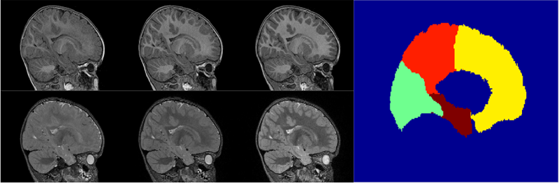
Modeling of early brain growth trajectories from longitudinal MRI will provide new insight into neurodevelopmental characteristics, timing and type of changes in neurological disorders from controls. In addition to an ongoing large-scale infant autism neuroimaging study 1, we recruited 4 infants with Down’s syndrome (DS) in order to evaluate newly developed methods for 4D segmentation from longitudinal infant MRI, and for temporal modeling of brain growth trajectories. Specifically to Down's, a comparison of patterns of full brain and lobar tissue growth may lead to better insight into the observed variability of cognitive development and neurological effects, and may help with development of disease-modifying therapeutic intervention.
Characterizing growth patterns in longitudinal {MRI} using image contrast
A. Vardhan, M. Prastawa, C. Vachet, J. Piven, G. Gerig.
“Characterizing growth patterns in longitudinal MRI using image contrast,” In Proceedings of Medical Imaging 2014: Image Processing, 2014.
ABSTRACT
×
Understanding the growth patterns of the early brain is crucial to the study of neuro-development. In the early stages of brain growth, a rapid sequence of biophysical and chemical processes take place. A crucial component of these processes, known as myelination, consists of the formation of a myelin sheath around a nerve fiber, enabling the effective transmission of neural impulses. As the brain undergoes myelination, there is a subsequent change in the contrast between gray matter and white matter as observed in MR scans. In this work, graywhite matter contrast is proposed as an effective measure of appearance which is relatively invariant to location, scanner type, and scanning conditions. To validate this, contrast is computed over various cortical regions for an adult human phantom. MR (Magnetic Resonance) images of the phantom were repeatedly generated using different scanners, and at different locations. Contrast displays less variability over changing conditions of scan compared to intensity-based measures, demonstrating that it is less dependent than intensity on external factors. Additionally, contrast is used to analyze longitudinal MR scans of the early brain, belonging to healthy controls and Down's Syndrome (DS) patients. Kernel regression is used to model subject-specific trajectories of contrast changing with time. Trajectories of contrast changing with time, as well as time-based biomarkers extracted from contrast modeling, show large differences between groups. The preliminary applications of contrast based analysis indicate its future potential to reveal new information not covered by conventional volumetric or deformation-based analysis, particularly for distinguishing between normal and abnormal growth patterns.
Joint Longitudinal Modeling of Brain Appearance in Multimodal {MRI} for the Characterization of Early Brain Developmental Processes
A. Vardhan, N. Sadeghi, C. Vachet, J. Piven, G. Gerig.
“Joint Longitudinal Modeling of Brain Appearance in Multimodal MRI for the Characterization of Early Brain Developmental Processes,” In Spatiotemporal Image Analysis for Longitudinal and Time-Series Image Data (STIA'14) , LNCS. MICCAI'14, Springer Verlag, June, 2014.
ABSTRACT
×
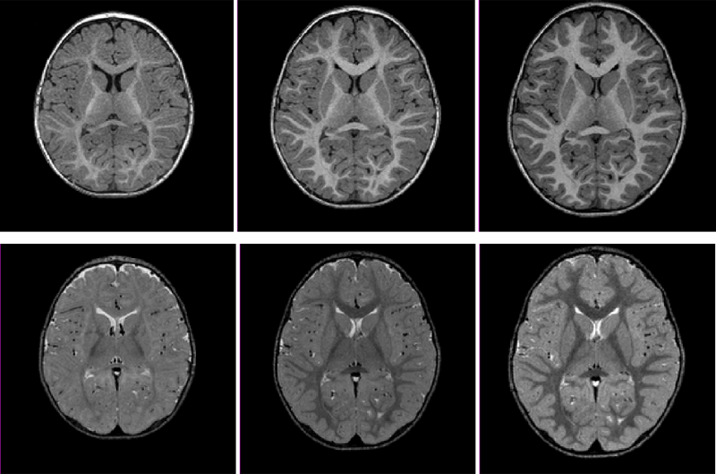
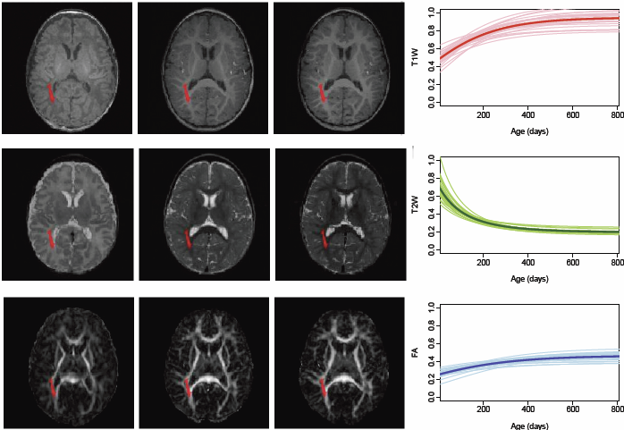
Early brain maturational processes such as myelination manifest as changes in the relative appearance of white-gray matter tissue classes in MR images. Imaging modalities such as T1W (T1-Weighted) and T2W (T2-Weighted) MRI each display specific patterns of appearance change associated with distinct neurobiological components of these maturational processes. In this paper we present a framework to jointly model multimodal appearance changes across time for a longitudinal imaging dataset, resulting in quantitative assessment of the patterns of early brain maturation not yet available to clinicians. We measure appearance by quantifying contrast between white and gray matter in terms of the distance between their intensity distributions, a method demonstrated to be relatively stable to interscan variability. A multivariate nonlinear mixed effects (NLME) model is used for joint statistical modeling of this contrast measure across multiple imaging modalities. The multivariate NLME procedure considers correlations between modalities in addition to intra-modal variability. The parameters of the logistic growth function used in NLME modeling provide useful quantitative information about the timing and progression of contrast change in multimodal datasets. Inverted patterns of relative white-gray matter intensity gradient that are observable in T1W scans with respect to T2W scans are characterized by the SIR (Signal Intensity Ratio). The CONTDIR (Contrast Direction) which measures the direction of the gradient at each time point relative to that in the adult-like scan adds a directional attribute to contrast. The major contribution of this paper is a framework for joint multimodal temporal modeling of white-gray matter MRI contrast change and estimation of subject-specific and population growth trajectories. Results confirm qualitative descriptions of growth patterns in pediatric radiology studies and our new quantitative modeling scheme has the potential to advance understanding of variability of brain tissue maturation and to eventually differentiate normal from abnormal growth for early diagnosis of pathology.
UNC-Utah NA-MIC framework for DTI fiber tract analysis
A.R. Verde, F. Budin, J.-B. Berger, A. Gupta, M. Farzinfar, A. Kaiser, M. Ahn, H. Johnson, J. Matsui, H.C. Hazlett, A. Sharma, C. Goodlett, Y. Shi, S. Gouttard, C. Vachet, J. Piven, H. Zhu, G. Gerig, M. Styner.
“UNC-Utah NA-MIC framework for DTI fiber tract analysis,” In Frontiers in Neuroinformatics, Vol. 7, No. 51, January, 2014.
DOI: 10.3389/fninf.2013.00051
ABSTRACT
×
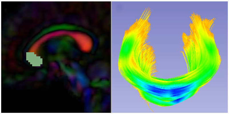
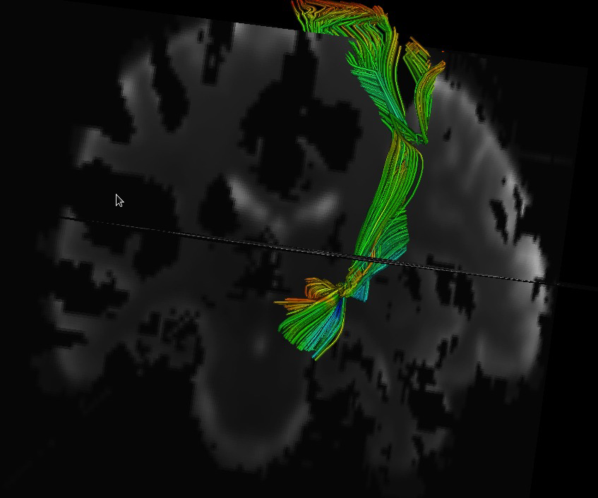
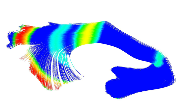
Diffusion tensor imaging has become an important modality in the field of neuroimaging to capture changes in micro-organization and to assess white matter integrity or development. While there exists a number of tractography toolsets, these usually lack tools for preprocessing or to analyze diffusion properties along the fiber tracts. Currently, the field is in critical need of a coherent end-to-end toolset for performing an along-fiber tract analysis, accessible to non-technical neuroimaging researchers. The UNC-Utah NA-MIC DTI framework represents a coherent, open source, end-to-end toolset for atlas fiber tract based DTI analysis encompassing DICOM data conversion, quality control, atlas building, fiber tractography, fiber parameterization, and statistical analysis of diffusion properties. Most steps utilize graphical user interfaces (GUI) to simplify interaction and provide an extensive DTI analysis framework for non-technical researchers/investigators. We illustrate the use of our framework on a small sample, cross sectional neuroimaging study of eight healthy 1-year-old children from the Infant Brain Imaging Study (IBIS) Network. In this limited test study, we illustrate the power of our method by quantifying the diffusion properties at 1 year of age on the genu and splenium fiber tracts.
Keywords: neonatal neuroimaging, white matter pathways, magnetic resonance imaging, diffusion tensor imaging, diffusion imaging quality control, DTI atlas building
Multi-atlas segmentation of subcortical brain structures via the AutoSeg software pipeline
J. Wang, C. Vachet, A. Rumple, S. Gouttard, C. Ouzie, E. Perrot, G. Du, X. Huang, G. Gerig, M.A. Styner.
“Multi-atlas segmentation of subcortical brain structures via the AutoSeg software pipeline,” In Frontiers in Neuroinformatics, Vol. 8, No. 7, 2014.
DOI: 10.3389/fninf.2014.00007
ABSTRACT
×
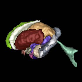
Automated segmenting and labeling of individual brain anatomical regions, in MRI are challenging, due to the issue of individual structural variability. Although atlas-based segmentation has shown its potential for both tissue and structure segmentation, due to the inherent natural variability as well as disease-related changes in MR appearance, a single atlas image is often inappropriate to represent the full population of datasets processed in a given neuroimaging study. As an alternative for the case of single atlas segmentation, the use of multiple atlases alongside label fusion techniques has been introduced using a set of individual “atlases” that encompasses the expected variability in the studied population. In our study, we proposed a multi-atlas segmentation scheme with a novel graph-based atlas selection technique. We first paired and co-registered all atlases and the subject MR scans. A directed graph with edge weights based on intensity and shape similarity between all MR scans is then computed. The set of neighboring templates is selected via clustering of the graph. Finally, weighted majority voting is employed to create the final segmentation over the selected atlases. This multi-atlas segmentation scheme is used to extend a single-atlas-based segmentation toolkit entitled AutoSeg, which is an open-source, extensible C++ based software pipeline employing BatchMake for its pipeline scripting, developed at the Neuro Image Research and Analysis Laboratories of the University of North Carolina at Chapel Hill. AutoSeg performs N4 intensity inhomogeneity correction, rigid registration to a common template space, automated brain tissue classification based skull-stripping, and the multi-atlas segmentation. The multi-atlas-based AutoSeg has been evaluated on subcortical structure segmentation with a testing dataset of 20 adult brain MRI scans and 15 atlas MRI scans. The AutoSeg achieved mean Dice coefficients of 81.73% for the subcortical structures.
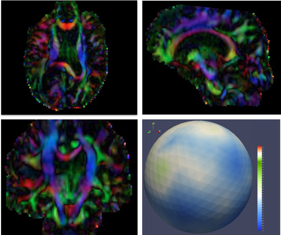
Diffusion MR imaging has received increasing attention in the neuroimaging community, as it yields new insights into the microstructural organization of white matter that are not available with conventional MRI techniques. While the technology has enormous potential, diffusion MRI suffers from a unique and complex set of image quality problems, limiting the sensitivity of studies and reducing the accuracy of findings. Furthermore, the acquisition time for diffusion MRI is longer than conventional MRI due to the need for multiple acquisitions to obtain directionally encoded Diffusion Weighted Images (DWI). This leads to increased motion artifacts, reduced signal-to-noise ratio (SNR), and increased proneness to a wide variety of artifacts, including eddy-current and motion artifacts, “venetian blind” artifacts, as well as slice-wise and gradient-wise inconsistencies. Such artifacts mandate stringent Quality Control (QC) schemes in the processing of diffusion MRI data. Most existing QC procedures are conducted in the DWI domain and/or on a voxel level, but our own experiments show that these methods often do not fully detect and eliminate certain types of artifacts, often only visible when investigating groups of DWI's or a derived diffusion model, such as the most-employed diffusion tensor imaging (DTI). Here, we propose a novel regional QC measure in the DTI domain that employs the entropy of the regional distribution of the principal directions (PD). The PD entropy quantifies the scattering and spread of the principal diffusion directions and is invariant to the patient's position in the scanner. High entropy value indicates that the PDs are distributed relatively uniformly, while low entropy value indicates the presence of clusters in the PD distribution. The novel QC measure is intended to complement the existing set of QC procedures by detecting and correcting residual artifacts. Such residual artifacts cause directional bias in the measured PD and here called dominant direction artifacts. Experiments show that our automatic method can reliably detect and potentially correct such artifacts, especially the ones caused by the vibrations of the scanner table during the scan. The results further indicate the usefulness of this method for general quality assessment in DTI studies.
Adaptive prior probability and spatial temporal intensity change estimation for segmentation of the one-year-old human brain
S.H. Kim, V. Fonov, C. Dietrich, C. Vachet, H.C. Hazlett, R.G. Smith, M. Graves, J. Piven, J.H. Gilmore, D.L. Collins, G. Gerig, M. Styner, The IBIS network.
“Adaptive prior probability and spatial temporal intensity change estimation for segmentation of the one-year-old human brain,” In Journal of Neuroscience Methods, Vol. 212, No. 1, Note: Published online Sept. 29, pp. 43--55. January, 2013.
DOI: 10.1016/j.jneumeth.2012.09.01
PubMed Central ID: PMC3513941
ABSTRACT
×
The degree of white matter (WM) myelination is rather inhomogeneous across the brain. White matter appears differently across the cortical lobes in MR images acquired during early postnatal development. Specifically at 1-year of age, the gray/white matter contrast of MR T1 and T2 weighted images in prefrontal and temporal lobes is reduced as compared to the rest of the brain, and thus, tissue segmentation results commonly show lower accuracy in these lobes. In this novel work, we propose the use of spatial intensity growth maps (IGM) for T1 and T2 weighted images to compensate for local appearance inhomogeneity. The IGM captures expected intensity changes from 1 to 2 years of age, as appearance homogeneity is greatly improved by the age of 24 months. The IGM was computed as the coefficient of a voxel-wise linear regression model between corresponding intensities at 1 and 2 years. The proposed IGM method revealed low regression values of 1–10\% in GM and CSF regions, as well as in WM regions at maturation stage of myelination at 1 year. However, in the prefrontal and temporal lobes we observed regression values of 20–25\%, indicating that the IGM appropriately captures the expected large intensity change in these lobes mainly due to myelination. The IGM is applied to cross-sectional MRI datasets of 1-year-old subjects via registration, correction and tissue segmentation of the IGM-corrected dataset. We validated our approach in a small leave-one-out study of images with known, manual 'ground truth' segmentations.
Lateral ventricle morphology analysis via mean latitude axis
B. Paniagua, A. Lyall, J.-B. Berger, C. Vachet, R.M. Hamer, S. Woolson, W. Lin, J. Gilmore, M. Styner.
“Lateral ventricle morphology analysis via mean latitude axis,” In Proceedings of SPIE 8672, Biomedical Applications in Molecular, Structural, and Functional Imaging, 86720M, 2013.
DOI: 10.1117/12.2006846
PubMed ID: 23606800
PubMed Central ID: PMC3630372
ABSTRACT
×
Statistical shape analysis has emerged as an insightful method for evaluating brain structures in neuroimaging studies, however most shape frameworks are surface based and thus directly depend on the quality of surface alignment. In contrast, medial descriptions employ thickness information as alignment-independent shape metric. We propose a joint framework that computes local medial thickness information via a mean latitude axis from the well-known spherical harmonic (SPHARM-PDM) shape framework. In this work, we applied SPHARM derived medial representations to the morphological analysis of lateral ventricles in neonates. Mild ventriculomegaly (MVM) subjects are compared to healthy controls to highlight the potential of the methodology. Lateral ventricles were obtained from MRI scans of neonates (9- 144 days of age) from 30 MVM subjects as well as age- and sex-matched normal controls (60 total). SPHARM-PDM shape analysis was extended to compute a mean latitude axis directly from the spherical parameterization. Local thickness and area was straightforwardly determined. MVM and healthy controls were compared using local MANOVA and compared with the traditional SPHARM-PDM analysis. Both surface and mean latitude axis findings differentiate successfully MVM and healthy lateral ventricle morphology. Lateral ventricles in MVM neonates show enlarged shapes in tail and head. Mean latitude axis is able to find significant differences all along the lateral ventricle shape, demonstrating that local thickness analysis provides significant insight over traditional SPHARM-PDM. This study is the first to precisely quantify 3D lateral ventricle morphology in MVM neonates using shape analysis.
Multivariate Modeling of Longitudinal {MRI} in Early Brain Development with Confidence Measures
N. Sadeghi, M.W. Prastawa, P.T. Fletcher, C. Vachet, Bo Wang, J.H. Gilmore, G. Gerig.
“Multivariate Modeling of Longitudinal MRI in Early Brain Development with Confidence Measures,” In Proceedings of the 2013 IEEE 10th International Symposium on Biomedical Imaging (ISBI), pp. 1400--1403. 2013.
DOI: 10.1109/ISBI.2013.6556795
ABSTRACT
×
The human brain undergoes rapid organization and structuring early in life. Longitudinal imaging enables the study of these changes over a developmental period within individuals through estimation of population growth trajectory and its variability. In this paper, we focus on maturation of white and gray matter as is depicted in structural and diffusion MRI of healthy subjects with repeated scans. We provide a framework for joint analysis of both structural MRI and DTI (Diffusion Tensor Imaging) using multivariate nonlinear mixed effect modeling of temporal changes. Our framework constructs normative growth models for all the modalities that take into account the correlation among the modalities and individuals, along with estimation of the variability of the population trends. We apply our method to study early brain development, and to our knowledge this is the first multimodel longitudinal modeling of diffusion and signal intensity changes for this growth stage. Results show the potential of our framework to study growth trajectories, as well as neurodevelopmental disorders through comparison against the constructed normative models of multimodal 4D MRI.
Analysis of Diffusion Tensor Imaging for Subjects with Down Syndrome
N. Sadeghi, C. Vachet, M. Prastawa, J. Korenberg, G. Gerig.
“Analysis of Diffusion Tensor Imaging for Subjects with Down Syndrome,” In Proceedings of the 19th Annual Meeting of the Organization for Human Brain Mapping OHBM, pp. (in print). 2013.
ABSTRACT
Down syndrome (DS) is the most common chromosome abnormality in humans. It is typically associated with delayed cognitive development and physical growth. DS is also associated with Alzheimer-like dementia [1]. In this study we analyze the white matter integrity of individuals with DS compared to control as is reflected in the diffusion parameters derived from Diffusion Tensor Imaging. DTI provides relevant information about the underlying tissue, which correlates with cognitive function [2]. We present a cross-sectional analysis of white matter tracts of subjects with DS compared to control.
Software-based diffusion MR human brain phantom for evaluating fiber-tracking algorithms
Y. Shi, G. Roger, C. Vachet, F. Budin, E. Maltbie, A. Verde, M. Hoogstoel, J.-B. Berger, M. Styner.
“Software-based diffusion MR human brain phantom for evaluating fiber-tracking algorithms,” In Proceedings of SPIE 8669, Medical Imaging 2013: Image Processing, 86692A, 2013.
DOI: 10.1117/12.2006113
PubMed ID: 24357914
PubMed Central ID: PMC3865235
ABSTRACT
×
Fiber tracking provides insights into the brain white matter network and has become more and more popular in diffusion magnetic resonance (MR) imaging. Hardware or software phantom provides an essential platform to investigate, validate and compare various tractography algorithms towards a "gold standard". Software phantoms excel due to their flexibility in varying imaging parameters, such as tissue composition, SNR, as well as potential to model various anatomies and pathologies. This paper describes a novel method in generating diffusion MR images with various imaging parameters from realistically appearing, individually varying brain anatomy based on predefined fiber tracts within a high-resolution human brain atlas. Specifically, joint, high resolution DWI and structural MRI brain atlases were constructed with images acquired from 6 healthy subjects (age 22-26) for the DWI data and 56 healthy subject (age 18-59) for the structural MRI data. Full brain fiber tracking was performed with filtered, two-tensor tractography in atlas space. A deformation field based principal component model from the structural MRI as well as unbiased atlas building was then employed to generate synthetic structural brain MR images that are individually varying. Atlas fiber tracts were accordingly warped into each synthetic brain anatomy. Diffusion MR images were finally computed from these warped tracts via a composite hindered and restricted model of diffusion with various imaging parameters for gradient directions, image resolution and SNR. Furthermore, an open-source program was developed to evaluate the fiber tracking results both qualitatively and quantitatively based on various similarity measures.
{UNC-Utah} {NA-MIC} {DTI} framework: atlas based fiber tract analysis with application to a study of nicotine smoking addiction
A.R. Verde, J.-B. Berger, A. Gupta, M. Farzinfar, A. Kaiser, V.W. Chanon, C. Boettiger, H. Johnson, J. Matsui, A. Sharma, C. Goodlett, Y. Shi, H. Zhu, G. Gerig, S. Gouttard, C. Vachet, M. Styner.
“UNC-Utah NA-MIC DTI framework: atlas based fiber tract analysis with application to a study of nicotine smoking addiction,” In Proc. SPIE 8669, Medical Imaging 2013: Image Processing, 86692D, Vol. 8669, pp. 86692D-86692D-8. 2013.
DOI: 10.1117/12.2007093
ABSTRACT
×
Purpose: The UNC-Utah NA-MIC DTI framework represents a coherent, open source, atlas fiber tract based DTI analysis framework that addresses the lack of a standardized fiber tract based DTI analysis workflow in the field. Most steps utilize graphical user interfaces (GUI) to simplify interaction and provide an extensive DTI analysis framework for non-technical researchers/investigators. Data: We illustrate the use of our framework on a 54 directional DWI neuroimaging study contrasting 15 Smokers and 14 Controls. Method(s): At the heart of the framework is a set of tools anchored around the multi-purpose image analysis platform 3D-Slicer. Several workflow steps are handled via external modules called from Slicer in order to provide an integrated approach. Our workflow starts with conversion from DICOM, followed by thorough automatic and interactive quality control (QC), which is a must for a good DTI study. Our framework is centered around a DTI atlas that is either provided as a template or computed directly as an unbiased average atlas from the study data via deformable atlas building. Fiber tracts are defined via interactive tractography and clustering on that atlas. DTI fiber profiles are extracted automatically using the atlas mapping information. These tract parameter profiles are then analyzed using our statistics toolbox (FADTTS). The statistical results are then mapped back on to the fiber bundles and visualized with 3D Slicer. Results: This framework provides a coherent set of tools for DTI quality control and analysis. Conclusions: This framework will provide the field with a uniform process for DTI quality control and analysis.
2012
Brain Volume Findings in Six Month Old Infants at High Familial Risk for Autism
H.C. Hazlett, H. Gu, R.C. McKinstry, D.W.W. Shaw, K.N. Botteron, S. Dager, M. Styner, C. Vachet, G. Gerig, S. Paterson, R.T. Schultz, A.M. Estes, A.C. Evans, J. Piven.
“Brain Volume Findings in Six Month Old Infants at High Familial Risk for Autism,” In American Journal of Psychiatry (AJP), pp. (in print). 2012.
ABSTRACT
Objective: Brain enlargement has been observed in individuals with autism as early as two years of age. Studies using head circumference suggest that brain enlargement is a postnatal event that occurs around the latter part of the first year. To date, no brain imaging studies have systematically examined the period prior to age two. In this study we examine MRI brain volume in six month olds at high familial risk for autism.
Method: The Infant Brain Imaging Study (IBIS) is a longitudinal imaging study of infants at high risk for autism. This cross-sectional analysis examines brain volumes at six months of age, in high risk infants (N=98) in comparison to infants without family members with autism (low risk) (N=36). MRI scans are also examined for radiologic abnormalities.
Results: No group differences were observed for intracranial cerebrum, cerebellum, lateral ventricle volumes, or head circumference.
Conclusions:We did not observe significant group differences for head circumference, brain volume, or abnormalities of radiologic findings in a sample of 6 month old infants at highrisk for autism. We are unable to conclude that these changes are not present in infants who later go on to receive a diagnosis of autism, but rather that they were not detected in a large group at high familial risk. Future longitudinal studies of the IBIS sample will examine whether brain volume may differ in those infants who go onto develop autism, estimating that approximately 20\% of this sample may be diagnosed with an autism spectrum disorder at age two.
Combined {SPHARM-PDM} and entropy-based particle systems shape analysis framework
B. Paniagua, L. Bompard, J. Cates, R.T. Whitaker, M. Datar, C. Vachet, M. Styner.
“Combined SPHARM-PDM and entropy-based particle systems shape analysis framework,” In Medical Imaging 2012: Biomedical Applications in Molecular, Structural, and Functional Imaging, SPIE Intl Soc Optical Eng, March, 2012.
DOI: 10.1117/12.911228
PubMed ID: 24027625
PubMed Central ID: PMC3766973
ABSTRACT
×
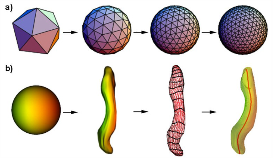
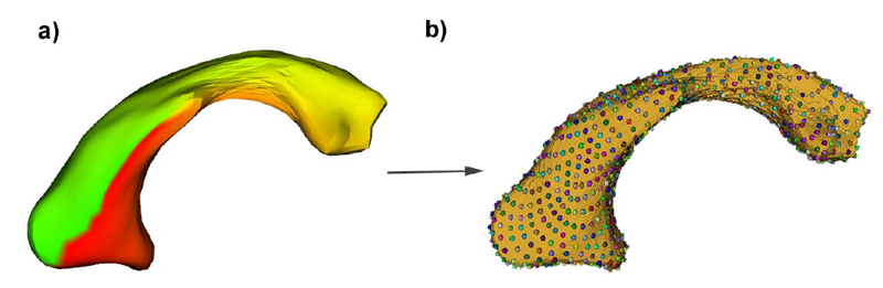
Description of purpose: The NA-MIC SPHARM-PDM Toolbox represents an automated set of tools for the computation of 3D structural statistical shape analysis. SPHARM-PDM solves the correspondence problem by defining a first order ellipsoid aligned, uniform spherical parameterization for each object with correspondence established at equivalently parameterized points. However, SPHARM correspondence has shown to be inadequate for some biological shapes that are not well described by a uniform spherical parameterization. Entropy-based particle systems compute correspondence by representing surfaces as discrete point sets that does not rely on any inherent parameterization. However, they are sensitive to initialization and have little ability to recover from initial errors. By combining both methodologies we compute reliable correspondences in topologically challenging biological shapes. Data: Diverse subcortical structures cohorts were used, obtained from MR brain images. Method(s): The SPHARM-PDM shape analysis toolbox was used to compute point based correspondent models that were then used as initializing particles for the entropy-based particle systems. The combined framework was implemented as a stand-alone Slicer3 module, which works as an end-to-end shape analysis module. Results: The combined SPHARM-PDM-Particle framework has demonstrated to improve correspondence in the example dataset over the conventional SPHARM-PDM toolbox. Conclusions: The work presented in this paper demonstrates a two-sided improvement for the scientific community, being able to 1) find good correspondences among spherically topological shapes, that can be used in many morphometry studies 2) offer an end-to-end solution that will facilitate the access to shape analysis framework to users without computer expertise.
Automatic corpus callosum segmentation using a deformable active Fourier contour model
C. Vachet, B. Yvernault, K. Bhatt, R.G. Smith, G. Gerig, H.C. Hazlett, M.A. Styner.
“Automatic corpus callosum segmentation using a deformable active Fourier contour model,” In Proceedings of Medical Imaging 2012: Biomedical Applications in Molecular, Structural, and Functional Imaging, SPIE, Vol. 8317, 831707, 2012.
DOI: 10.1117/12.911504
ABSTRACT
×
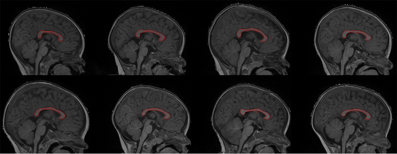
The corpus callosum (CC) is a structure of interest in many neuroimaging studies of neuro-developmental pathology such as autism. It plays an integral role in relaying sensory, motor and cognitive information from homologous regions in both hemispheres.
We have developed a framework that allows automatic segmentation of the corpus callosum and its lobar subdivisions. Our approach employs constrained elastic deformation of exible Fourier contour model, and is an extension of Szekely's 2D Fourier descriptor based Active Shape Model. The shape and appearance model, derived from a large mixed population of 150+ subjects, is described with complex Fourier descriptors in a principal component shape space. Using MNI space aligned T1w MRI data, the CC segmentation is initialized on the mid-sagittal plane using the tissue segmentation. A multi-step optimization strategy, with two constrained steps and a final unconstrained step, is then applied. If needed, interactive segmentation can be performed via contour repulsion points. Lobar connectivity based parcellation of the corpus callosum can finally be computed via the use of a probabilistic CC subdivision model.
Our analysis framework has been integrated in an open-source, end-to-end application called CCSeg both with a command line and Qt-based graphical user interface (available on NITRC). A study has been performed to quantify the reliability of the semi-automatic segmentation on a small pediatric dataset. Using 5 subjects randomly segmented 3 times by two experts, the intra-class correlation coeficient showed a superb reliability (0.99). CCSeg is currently applied to a large longitudinal pediatric study of brain development in autism.
2011
Early Brain Overgrowth in Autism Associated with an Increase in Cortical Surface Area Before Age 2
H.C. Hazlett, M. Poe, G. Gerig, M. Styner, C. Chappell, R.G. Smith, C. Vachet, J. Piven.
“Early Brain Overgrowth in Autism Associated with an Increase in Cortical Surface Area Before Age 2,” In Arch of Gen Psych, Vol. 68, No. 5, pp. 467--476. 2011.
DOI: 10.1001/archgenpsychiatry.2011.39
Spatial Intensity Prior Correction for Tissue Segmentation in the Developing human Brain
S.H. Kim, V. Fonov, J. Piven, J. Gilmore, C. Vachet, G. Gerig, D.L. Collins, M. Styner.
“Spatial Intensity Prior Correction for Tissue Segmentation in the Developing human Brain,” In Proceedings of IEEE ISBI 2011, pp. 2049--2052. 2011.
DOI: 10.1109/ISBI.2011.5872815
2010
Changes of MR and DTI appearance in early human brain development
C. Marc, C. Vachet, J.E. Blocher, G. Gerig, J.H. Gilmore, M.A. Styner.
“Changes of MR and DTI appearance in early human brain development,” In Proceedings of SPIE Medical Imaging 7623, 762324, 2010.
DOI: 10.1117/12.844912
2009
Cortical Correspondence with Probabilistic Fiber Connectivity
I. Oguz, M. Niethammer, J. Cates, R.T. Whitaker, P.T. Fletcher, C. Vachet, M. Styner.
“Cortical Correspondence with Probabilistic Fiber Connectivity,” In Information Processing in Medical Imaging (IPMI), Lecture Notes in Computer Science (LCNS), Vol. 5636, pp. 651--663. 2009.
DOI: 10.1007/978-3-642-02498-6_54