
SCI Publications
2009
![]()
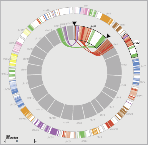
M.D. Meyer, T. Munzner, H. Pfister.
“MizBee: A Multiscale Synteny Browser,” In IEEE Transactions on Visualization and Computer Graphics (Proceedings of InfoVis 2009), Vol. 15, No. 6, Note: Honorable Mention for Best Paper Award, pp. 897--904. 2009.
![]()
M. Milanic, V. Jazbinsek, D.F. Wang, J. Sinstra, R.S. Macleod, D.H. Brooks, R. Hren.
“Evaluation of Approaches of Solving Electrocardiographic Imaging Problem,” In Proceeding of Computers in Cardiology 2010, Park City, Utah, September, 2009.
![]()
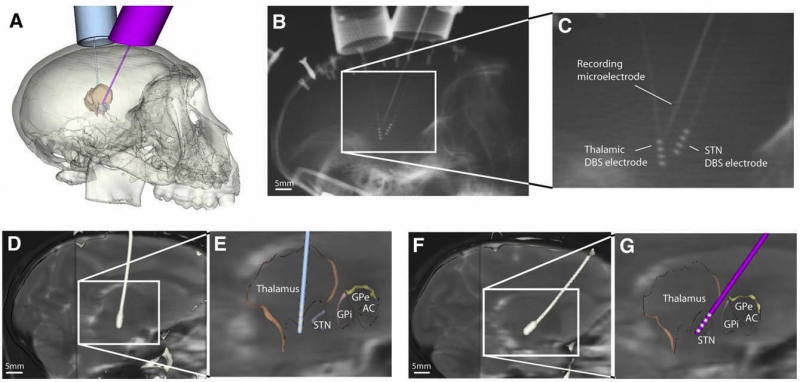
S. Miocinovic, S.F. Lempka, G.S. Russo, C.B. Maks, C.R. Butson, K.E. Sakaie, J.L. Vitek, C.C. McIntyre.
“Experimental and theoretical characterization of the voltage distribution generated by deep brain stimulation,” In Experimental Neurology, Vol. 216, No. 1, Elsevier Inc., pp. 166--176. March, 2009.
ISSN: 1090--2430
DOI: 10.1016/j.expneurol.2008.11.024
PubMed ID: 19118551
![]()
H. Mirzaee, C. Eskilsson, S.J. Sherwin, R.M. Kirby.
“Comparison of Consistent Integration Versus Adaptive Quadrature for Taming Aliasing Errors,” SCI Technical Report, No. UUSCI-2009-008, SCI Institute, University of Utah, 2009.
![]()
S.M. Moore, B.J. Ellis, J.A. Weiss, P.J. McMahon, R.E. Debski.
“The Glenohumeral Capsule Should be Evaluated as a Sheet of Fibrous Tissue: A Validated Finite Element Model,” In Annals of Biomedical Engineering, Vol. 38, No. 1, pp. 66--76. 2009.
![]()
M.W. Mosconi, H. Cody-Hazlett, M.D. Poe, G. Gerig, R. Gimpel-Smith, J. Piven.
“Longitudinal study of amygdala volume and joint attention in 2- to 4-year-old children with autism,” In Arch Gen Psychiatry, Vol. 66, No. 5, pp. 509--516. 2009.
PubMed ID: 19414710
![]()
N. Mukherjee, C. Kang, H.M. Wolfe, B.S. Hertzberg, J.K. Smith, W. Lin, G. Gerig, R.M. Hamer, J.H. Gilmore.
“Discordance of Prenatal and Neonatal Brain Development in Twins,” In Early Human Development, Vol. 85, No. 3, pp. 171--175. 2009.
PubMed ID: 18804925
![]()
H Müller, R Reihs, S Sauer, K Zatloukal, M Streit, A Lex, B Schlegl, D Schmalstieg.
“Connecting Genes with Diseases,” In Information Visualisation, 2009 13th International Conference, pp. 323--330. July, 2009.
DOI: 10.1109/IV.2009.86
This paper presents a visual data mining approach using the combination of clinical data, pathways and gene-expression data. The visual exploration of medical data using pathways to navigate and filter the data allows a more systematic and efficient investigation of problems in modern life science. A multiplicity of hypothesis can be evaluated in the same period of time, enabling a much better exploitation of the data. We present a system for data preprocessing and automatic classification, a set of visualization views and finally the integration in the Caleydo visualization framework, which enables the "coupling" of genetic and a broad spectrum of clinical data. With the help of the Caleydo framework the medical expert can identify connections between genetic parameters, patient subgroups, and drug responses.
![]()
R.S. Oakes, T.J. Badger, E.G. Kholmovski, N. Akoum, N.S. Burgon, E.N. Fish, J.J. Blauer, S.N. Rao, E.V. DiBella, N.M. Segerson, M. Daccarett, J. Windfelder, C.J. McGann, D.L. Parker, R.S. MacLeod, N.F. Marrouche.
“Detection and quantification of left atrial structural remodeling with delayed-enhancement magnetic resonance imaging in patients with atrial fibrillation,” In Circulation, Vol. 119, No. 13, pp. 1758--1767. 2009.
![]()
I. Oguz, M. Niethammer, J. Cates, R.T. Whitaker, P.T. Fletcher, C. Vachet, M. Styner.
“Cortical Correspondence with Probabilistic Fiber Connectivity,” In Information Processing in Medical Imaging (IPMI), Lecture Notes in Computer Science (LCNS), Vol. 5636, pp. 651--663. 2009.
DOI: 10.1007/978-3-642-02498-6_54
![]()
![]()
L. Omberg, J.R. Meyerson, K. Kobayashi, L.S. Drury, J.F.X. Diffley, O. Alter.
“Global Effects of DNA Replication and DNA Replication Origin Activity on Eukaryotic Gene Expression,” In Nature Molecular Systems Biology, Vol. 5, No. 312, pp. (published online). October, 2009.
DOI: 10.1038/msb.2009.70
![]()
A.R.C. Paiva, I. Park, J.C. Principe.
“A Reproducing Kernel Hilbert Space Framework for Spike Train Signal Processing,” In Neural Computation, Vol. 21, No. 2, pp. 424--449. 2009.
![]()
Y. Pan, R.T. Whitaker, A. Cheryauka, D. Ferguson.
“Feasibility of GPU-Assisted Iterative Image Reconstruction for Mobile C-ARM CT,” In Progress in biomedical optics and imaging, In Proceedings of SPIE Medical Imaging 2009, Vol. 10, No. 3, 2009.
ISSN: 1605-7422
Computed tomography (CT) has been extensively studied and widely used for a variety of medical applications. The reconstruction of 3D images from a projection series is an important aspect of the modality. Reconstruction by filtered backprojection (FBP) is used by most manufacturers because of speed, ease of implementation, and relatively few parameters. Iterative reconstruction methods have a significant potential to provide superior performance with incomplete or noisy data, or with less than ideal geometries, such as cone-beam systems. However, iterative methods have a high computational cost, and regularization is usually required to reduce the effects of noise. The simultaneous algebraic reconstruction technique (SART) is studied in this paper, where the Feldkamp method (FDK) for filtered back projection is used as an initialization for iterative SART. Additionally, graphics hardware is utilized to increase the speed of SART implementation. Nvidia processors and compute unified device architecture (CUDA) form the platform for GPU computation. Total variation (TV) minimization is applied for the regularization of SART results. Preliminary results of SART on 3-D Shepp-Logan phantom using using TV regularization and GPU computation are presented in this paper. Potential improvements of the proposed framework are also discussed.
![]()
Y. Pan, W.-K. Jeong, R.T. Whitaker.
“Markov Surfaces: A Probabilistic Framework for User-Assisted Three Dimensional Image Segmentation,” In Proceedings of MICCAI 2009 workshop on Probabilistic Models for Medical Image Analysis, London, UK, 2009.
![]()
S.G. Parker, K. Damevski, A. Khan, A. Swaminathan, C.R. Johnson.
“The SCIJump Framework for Parallel and Distributed Scientific Computing,” In Advanced Computational Infrastructures for Parallel and Distributed Adaptive Applications, Edited by Manish Parashar and Xiaolin Li and Sumir Chandra, Wiley-Blackwell, pp. 149--170. 2009.
DOI: 10.1002/9780470558027.ch9
![]()
V. Pegoraro, S.G. Parker.
“An Analytical Solution to Single Scattering in Homogeneous Participating Media,” In Computer Graphics Forum (Proceedings of the 30th Eurographics Conference), Vol. 28, No. 2, pp. 329--335. 2009.
![]()
V. Pegoraro, M. Schott, S.G. Parker.
“An Analytical Approach to Single Scattering for Anisotropic Media and Light Distributions,” In Proceedings of the 35th Graphics Interface Conference, pp. 71--77. 2009.
![]()
N.S. Phatak, S.A. Maas, A.I. Veress, N.A. Pack, E.V. DiBella, J.A. Weiss.
“Strain Measurement in the Left Ventricle During Systole with Deformable Image Registration,” In Medical Image Analysi, Vol. 13, No. 2, pp. 354--361. 2009.
![]()
K. Potter, A. Wilson, P.-T. Bremer, D. Williams, C. Doutriaux, V. Pascucci, C.R. Johhson.
“Visualization of Uncertainty and Ensemble Data: Exploration of Climate Modeling and Weather Forecast Data with Integrated ViSUS-CDAT Systems,” In J. Phys.: Conf. Ser., Vol. 180, No. 012089, IOP Publishing, pp. 012089. July, 2009.
DOI: 10.1088/1742-6596/180/1/012089
![]()
K. Potter, A. Wilson, P.-T. Bremer, D. Williams, C. Doutriaux, V. Pascucci, C.R. Johnson.
“Ensemble-Vis: A Framework for the Statistical Visualization of Ensemble Data,” In Proceedings of the 2009 IEEE International Conference on Data Mining Workshops, pp. 233--240. 2009.
Page 74 of 144