 |
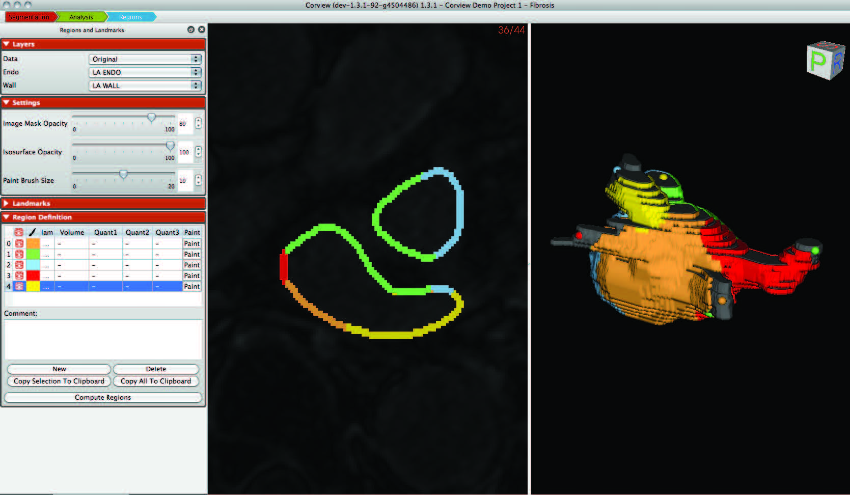
| Corview screenshot |
An interdisciplinary team at the Comprehensive Arrhythmia and MAnagement (CARMA) Center have made use of the segmentation, image analysis, and recently mesh generation and simulation capabilities of the CIBC to create a comprehensive program for AF management. The scope of the progress continues to expand each year and this application of CIBC technology has proven very fruitful even as it is very challenging.
The focus on this particular highlight is the unique use of the CIBC Seg3D software as not only a template but also an integral part of a dedicated, stand-alone workflow. The redesign of Seg3D that we have reported in the past had as a primary goal the capacity to separate the user interface from the core functionality and then share that functionality with other programs. This is precisely what has occurred with a the program Corview, which the CARMA center has developed from Seg3D. Corview is now the main workhorse of the entire image processing system at the CARMA Center and is used daily by 5-10 image analysis experts to extract from MRI scans from AF patients the information relevant for diagnosis, therapy planning, and clinical research. The entire workflow is on the verge of commercialization and we hope to reveal details of the arrangement very soon.
Atrial Fibrillation and Fibrosis
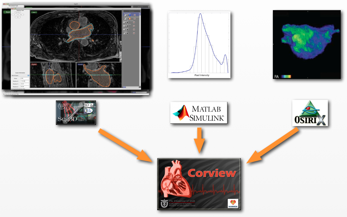
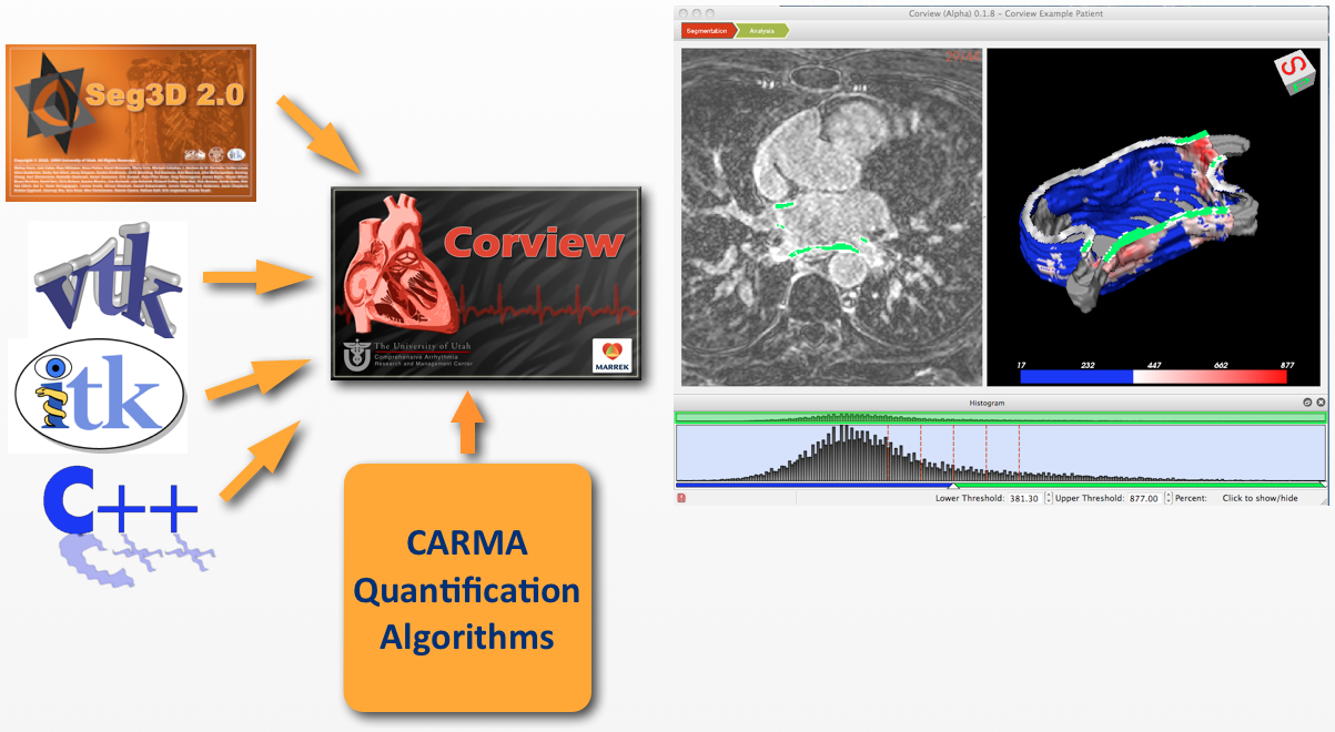
The fact that a relationship exists between increased fibrosis in the left atrium and the condition of atrial fibrillation (AF) is well established. However, until recently there was no way to capture the state of fibrosis in patients. Moreover, the precise mechanisms linking fibrosis to AF remain unknown and a topic of intense research around the world. The CARMA group, with assistance from the CIBC, has developed first a specialized MRI acquisition protocol and then a set of tools to extract from those images both structural and tissue substrate information.The figures in the left indicates the workflow schematically and also describes the discrete tools used in the past for this task. The use of separate tools, while relatively quick to implement and flexible to adjust, results in a routine that lacks integration and is also slow and prone to error compared to a more integrated approach. The second figure captures the more integrated workflow and the process by which Corview was created through merging a set of tools, almost all based on open source and commonly available modules. Corview contains all the elements of the old workflow but allowed a task that took hours to now minutes. Moreover, the infrastructure provided by Corview has enabled ongoing extension of its capabilities, meeting the ever changing needs of the CARMA group.
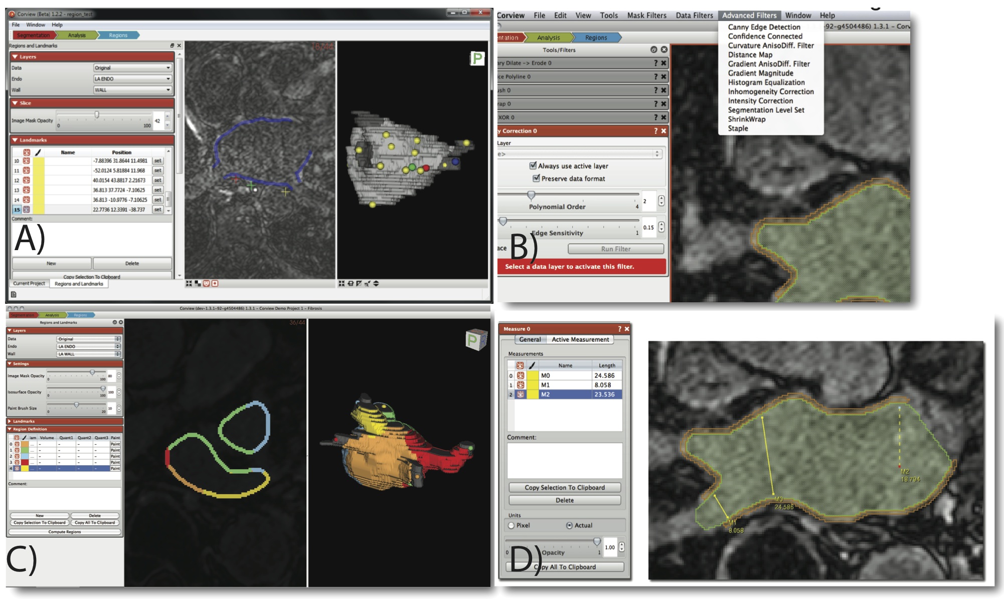
The image at the bottom of this page captures four of the feature sets that CARMA developers have created based on the infrastructure described above. With Corview, it is now possible to set and manipulate landmarks, essential elements for registration of images across patients and across time. The user can also select from a wide range of image filters, each based on an implementation of an Insight Toolkit function. And for quantification of tissue features, there is support for discrete measurements of distance, areas, and volumes as well as for regional quantification of fibrosis or scar-based on elements of the heart anatomy that the use can select interactively.


