Down syndrome is a common chromosomal disorder that affects 0.15 percent of the total population. Individuals with Down syndrome are prone to spinal instability due to congenital abnormalities in the occipital-cervical (O-C1) joint near the base of the skull. To determine the possible abnormality causing this instability, an image-processing pipeline was created by combining several available software packages and custom made software. Patients with Down syndrome and spinal instability at the O-C1 joint were age-matched with controls. The subject data was assessed and a congenital abnormality was defined in the superior articular facets of C1 in the Down syndrome patients. The software developed helped to visualize the abnormality and could be used in a clinical setting to help aide in the diagnosis and screening for spinal instability in Down syndrome patients.
Methods
To create a three dimensional model and determine the possible cause of O-C1 instability in Down syndrome patients, the following methods were employed:- Edge preserving and noise reducing data filtration
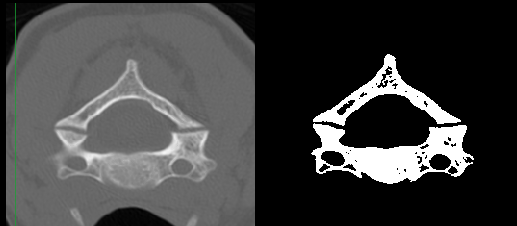
- Region Growing Segmentation multiple times on each data set (Fig. 1)
- Combination of Segmentations for each data set1
- Calculation of the tangent between the up vector and normal at each point in the volume
- Anti-aliasing filtration2
- Rendering2
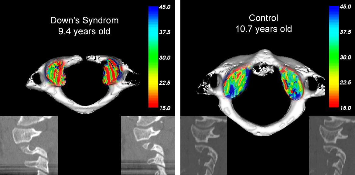
- Color mapping2 (Fig. 2)
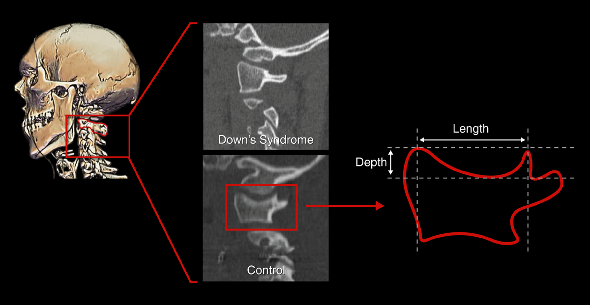
- Morphometric analysis from length and depth measurements along the midline of the lateral mass for each facet (Fig. 3)
 Fig. 1. Image before and after region growing segmentation. Fig. 1. Image before and after region growing segmentation. |
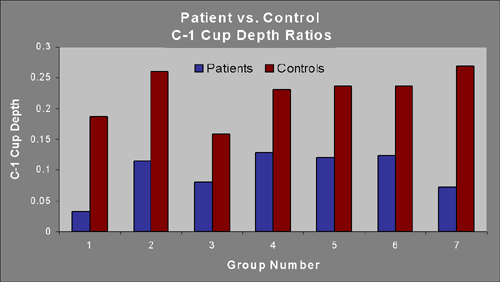
 Fig. 3. Morphometric analysis of the first cervical vertebral body was preformed by creating a normalization ratio of the depth over the length for each facet in the control and patient groups. Fig. 3. Morphometric analysis of the first cervical vertebral body was preformed by creating a normalization ratio of the depth over the length for each facet in the control and patient groups. |
|
 Table 2. Shows patient group normalization ratio compared to the control group normalization ratios. Table 2. Shows patient group normalization ratio compared to the control group normalization ratios. |
Discussion and Conclusions
Overall, by segmenting the images and rendering them with a color map, it was demonstrated that there was a difference in slope between the Down syndrome patients and the age matched controls. In addition, morphometric and statistical analysis show a significant difference between the control and Down patient groups. In the future, this application could be used to help assess spinal instability in children prior to injury.References
Special thanks to Samuel Browd MD, PhD, and Douglas Brockmeyer MD for the medical consultation, collaboration, and the images they provided for this study.1 Ibanez, L., Schroeder, W., Ng, L., Cates, J. The ITK Software Guide: The Insight Segmentation and Registration Toolkit (version 1.4). Albany: Kitware Inc., 2003.
2 Schroeder, W. J., K. M. Martin, B. E. Lorensen. The Visualization Toolkit, Third Edition. Albany: Kitware Inc., 2003.

