Tolga Tasdizen to Serve as a USTAR Faculty Member Within the SCI Institute
 Dr. Tolga Tasdizen has joined the SCI faculty as an Assistant Professor of the Department of Electrical and Computer Engineering and will become the SCI Institute's second USTAR Faculty member, joining Dr. Guido Gerig. Prior to being appointed USTAR Faculty, Tolga was a Research Assistant Professor in the School of Computing at the University of Utah. Dr. Tasdizen received his Ph.D. in Engineering from Brown University and is an expert in image processing, creating state-of-the-art research applied to biomedical and biological applications such as reconstructing neural circuit diagrams from large numbers of very high resolution microscopy images. His research interests include image analysis, computer vision and pattern recognition. The Utah Science and Technology Research Initiative (USTAR) is a state-funded, long-term effort to strengthen Utah's "knowledge economy".
Steve Corbató Named Director of Cyberinfrastructure Strategic Initiatives at the University of Utah
 The Scientific Computing and Imaging Institute congratulates Dr. Steve Corbató on being named Director of Cyberinfrastructure Strategic Initiatives at the University of Utah. Dr. Corbató has served as the SCI Institute's Associate director since May of 2006. Steve will report directly to VP of Information Technology, Steve Hess and help the University with future cyberinfrastructure plans. While Steve will be moving to the INSCC Building, we'll definitely still see him on a regular basis as we'll work together on future computing initiatives. Steve is already working on multiple projects and ideas with SCI Institute and other University of Utah faculty.
Center for Computational Earth Sciences at the SCI Institute
 Ross Whitaker (PI), Chuck Hansen, Claudio Silva, Valerio Pascucci, and Greg Jones have been awarded funding to create the Center for Computational Earth Sciences at the SCI Institute.
Visualizing Election Polls
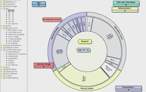 A New, Animated, Interactive Way to Analyze Opinion Data
Media Contacts
Oct. 6, 2008 - Do you want to know the percentage of white women who support vice presidential candidate Sarah Palin? What about college-educated versus high school-educated white women? Or those who also hunt?
University of Utah computer scientists have written software they hope eventually will allow news reporters and citizens to easily, interactively and visually answer such questions when analyzing election results, political opinion polls or other surveys.
NVIDIA Recognizes University Of Utah as a CUDA Center Of Excellence
 University of Utah Latest in a Growing List of Exceptional Schools Demonstrating Pioneering Work in Parallel Computing
Santa Clara, CA & Salt Lake City, UT - July 31, 2008 - NVIDIA Corporation, the worldwide leader in visual computing technologies, and the University of Utah today announced that the university has been recognized as a CUDA Center of Excellence, a milestone that marks the beginning of a significant partnership between the two organizations.
CRCNS: Fighting Blindness
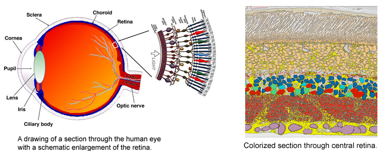
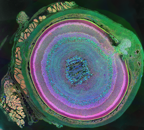 Despite great advances in neuroscience and medical technology in recent decades, nearly ten million Americans still suffer blindness due to retinal degenerative diseases such as retinitis pigmentosa (RP), age-related macular degeneration (AMD), diabetic retinopathy, and glaucoma. Unfortunately, current treatments available for these conditions are still quite limited. A primary challenge to developing effective treatments is the need for a complete understanding of the highly complex and delicate systems that compose the retina and how those systems change in response to degenerative disorders.
Remodeling processes that occur in the neuronal pathways within the retina during the course of retinal deterioration are of particular importance to the development of treatments for these conditions. Researchers at the Robert E. Marc Laboratory at the Moran Eye Center are collaborating with the SCI Institute on a project supported by the NIH-NIBIB (grant number 5R01EB005832) to develop high-throughput techniques for reconstructing and visualizing the neural structures that compose the retina in order to meet these challenges.
Scientific Background
Remembering Gene Golub - Salt Lake City, Utah
Those attending the event in Salt Lake City were:
Nelson Beebe, Department of Mathematics, University of Utah
Adam Bargteil - Carnegie Mellon University
Martin Berzins, School of Computing and SCI Institute, University of Utah
Mary Anne Berzins, Human Resources, University of Utah
Elaine Cohen, School of Computing, University of Utah
Kate Coles, Department of English, University of Utah
Steve Corbato, Office of Information Technology, University of Utah
Chuck Hansen, School of Computing and SCI Institute, University of Utah
Chris Johnson, School of Computing and SCI Institute, University of Utah
Greg Jones, SCI Institute
Tom Lyche, Department of Informatics, University of Oslo
Rich Riesenfeld, School of Computing, University of Utah
Kris Sikorski, School of Computing, University of Utah
Claudio Silva, School of Computing and SCI Institute, University of Utah
Barry Weller (Gene's Cousin), Department of English, University of Utah
Autism Research Profiled in Salt Lake Magazine
 The February 2008 Issue of Salt Lake magazine includes a profile of groundbreaking research being conducted at the University of Utah on the problem of Autism. Advancements in brain image analysis techniques developed by SCI researchers Guido Gerig, Ross Whitaker and P. Thomas Fletcher are specifically mentioned. (print version only)
Guido Gerig Joins Scientific Computing and Imaging Institute
 Guido Gerig joins the University of Utah's Scientific Computing and Imaging (SCI) Institute as a USTAR faculty member. The Scientific Computing and Imaging (SCI) Institute has established itself as an international research leader in the areas of scientific computing, scientific visualization, and image processing. USTAR is an innovative, aggressive and far-reaching effort to bolster Utah's economy with high-paying jobs and keep the state vibrant in the Knowledge Age. The USTAR Support Coalition and the Salt Lake Chamber sought public and private investment to recruit world-class research teams in carefully targeted disciplines. These teams will develop products and services that can be commercialized in new businesses and industries.
Neuro Image Analysis Team Joins SCI Institute
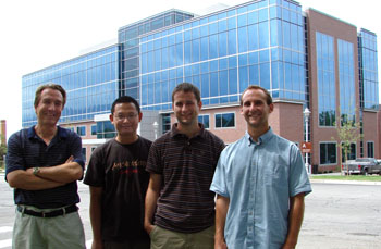 (L-R) Dr. Guido Gerig, Dr. Marcel Prastawa, Casey Goodlett, Sylvain Gouttard |

