
SCI Institute Appeals to Local Business Leadership for Increases In State Funding
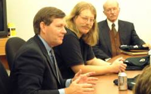
 On January 17th Dr. Christopher Johnson, director of the SCI Institute, appealed to local information technology business leaders to assist in lobbying the Utah State Legislature for more funding for Engineering disciplines. Dr. Johnson presented an overview of the current research within the Institute with an emphasis on medicine.
He described the history of biomedical devices from EEG to MRI. The talk also included the story of a real patient treated in part with technology developed here at the SCI Institute. Dr. Johnson described the case of Sarah, a child diagnosed with a tumor near the top of her spinal column. In such cases, doctors are only able to see sets of two-dimensional slices generated from an MRI. The physician must then reconstruct a three-dimensional image from those slices. Using technology developed at the SCI Institute, the team of physicians was not only able to see the tumor in full three-dimensions, but they were also able to use stereo glasses to “virtually” maneuver inside of the MRI. The combination of these technologies gave the surgeons a much clearer visualization and persuaded them to alter how they were going to operate.
SCI Institute Hosts Governor to Launch Ambitious Engineering Educational Initiative
 On March 19th, Governor Michael Leavitt visited the SCI Institute for a bill signing ceremony that marks the launch of an ambitious initiative to double the number of engineering and technology graduates over the next five years. The law provides funding for new faculty, pay increases, student loans, and infrastructure improvements. Most notably, it provides $15 million in matching funds for the construction of a new engineering building as well as $5 million for remodeling of the current Merrill Engineering Building. The new building will go directly north of the MEB. Gerald Stringfellow, the dean of the U of U College of Engineering, will be challenged to raise an additional $13 million in order to get the funding. Surrounded by students and some of the state's top political leaders, Gov. Leavitt discussed the importance of the new law. “This initiative is an essential step toward accelerating Utah's growth as a technology center,” he said. “It will help employers who face a critical shortage of engineers, and it will provide high-paying jobs for our children.”
SCI Institute Director Gives Plenary Talk on Visualization at Supercomputing 2001
 Dr. Chris Johnson delivered a plenary talk entitled "Scientific Visualization: Bridging the Complexity Threshold" at this year's Supercomputing conference in Denver, Colorado. Dr. Johnson discussed the need to bridge the "complexity threshold" in order to develop useful applications for analyzing data. He reviewed the state-of-the-art in visualization techniques, their use in real world applications, and presented an outline for future visualization research.
BISTI Grant Awarded to SCI
 The SCI Institute has been formally awarded a National Institutes of Health grant from the Biomedical Information Science and Technology Initiative(BISTI). This grant will be used to create a Program of Excellence in Computational Bioimaging and Visualization at the University of Utah. The award totals $2,261,136, distributed over three years, and includes collaborators from the departments of Bioengineering, Radiology, Neurosurgery and the Cardiovascular Research and Training Institute (CVRTI). Through awards such as these, NIH Institutes and Centers seek to establish National Programs of Excellence in Biomedical Computing (NPEBC). Funding for the SCI Institute grant comes from the National Heart, Lung, and Blood Institute within the National Institutes of Health.
SCI and the Governor's New Ecosystem
 On August 28, 2001, Dr. Chris Johnson attended a press conference conducted by Utah Governor Mike Leavitt. The governor launched a new program to generate business activity and investment in Utah's high technology ecosystems. Each ecosystem consists of academic researchers, business leaders, anchor companies, venture capitalists, and professional service providers who collectively promote business activity. Dr. Johnson spoke at this press conference with regard to the multidisciplinary nature of ongoing research at the SCI Institute.
SCI Institute Establishes Access Grid Node
 With the rapid advancement of new communication technologies, today's scientists are able to communicate more frequently and easily with collaborators and colleagues throughout the world. Collaborations used to require travelling on sabbaticals and meeting at infrequent conferences. Today, researchers need a way to collaborate remotely beyond the constraints of email and FTP.
Today's Tools - Tomorrow's Scientists: An ImageVis3D Project
 The SCI Institute holds a strong belief that providing research opportunities to undergraduate and even high school students will not only encourage them to pursue studies in the sciences, but also give them a head start in their future academic lives. Being allowed to work side by side with PhD-level scientists within a real research institute moves science from something that happens in a text book or highly structured laboratory to the dynamic work environment shared by scientists around the world.
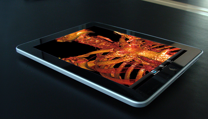
In this year's high school summer intern program, the SCI Institute invited four students, one each from Juan Diego Catholic High School, The Waterford School, and two from West High School. These students were given the opportunity to work with a lead software developer from the National Institutes of Health (NIH) sponsored Center for Integrative Biomedical Computing (CIBC). Their task seemed simple: take Seg3D and ImageVis3D (two advanced software tools developed by the CIBC), find a dataset of interest to the student, load that data, and experiment with the software on both desktop and iPad versions. And then, present your results to your high school peers. In the end, the students learned that research is a full-contact sport, not just a homework assignment. They had to 'dig-in', expand their knowledge, and learn about their subjects of interest, their data, their software, even their computers. In the end, the students translated this process and knowledge to science classes at their school. And, the top question after the presentations? Oddly enough, "how do I get an internship like yours?" Kids excited about a science internship! Mission Accomplished.
Mobile Mayhem: Researchers Harness Kraken to Model Explosions via Transport
by Gregory Scott Jones - NICS
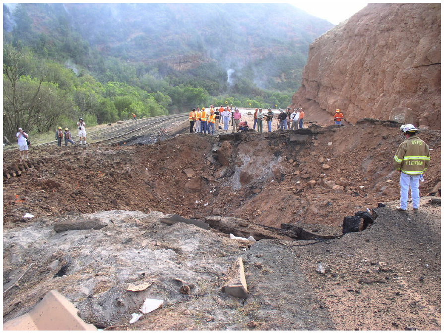 |
| The crater resulting from the Spanish Fork Detonation. |
Now, the good news: America's track record in transporting these materials is about as safe as they come. Very rarely, almost never in fact, are the potential dangers of these transports realized, largely due to instituted safeguards that seem to work very well.
However, accidents can happen. Take the August 2005 incident in Spanish Fork Canyon, Utah, for instance. A truck carrying 35,500 pounds of explosives—specifically small boosters used in seismic testing—overturned and exploded, creating a crater in the highway estimated to be between 20 to 35 feet deep and 70 feet wide according to the Utah Department of Transportation. But the damage wasn't solely financial. Four people, including the truck driver and a passenger, were hospitalized.
Neural Bioelectricity with EGI
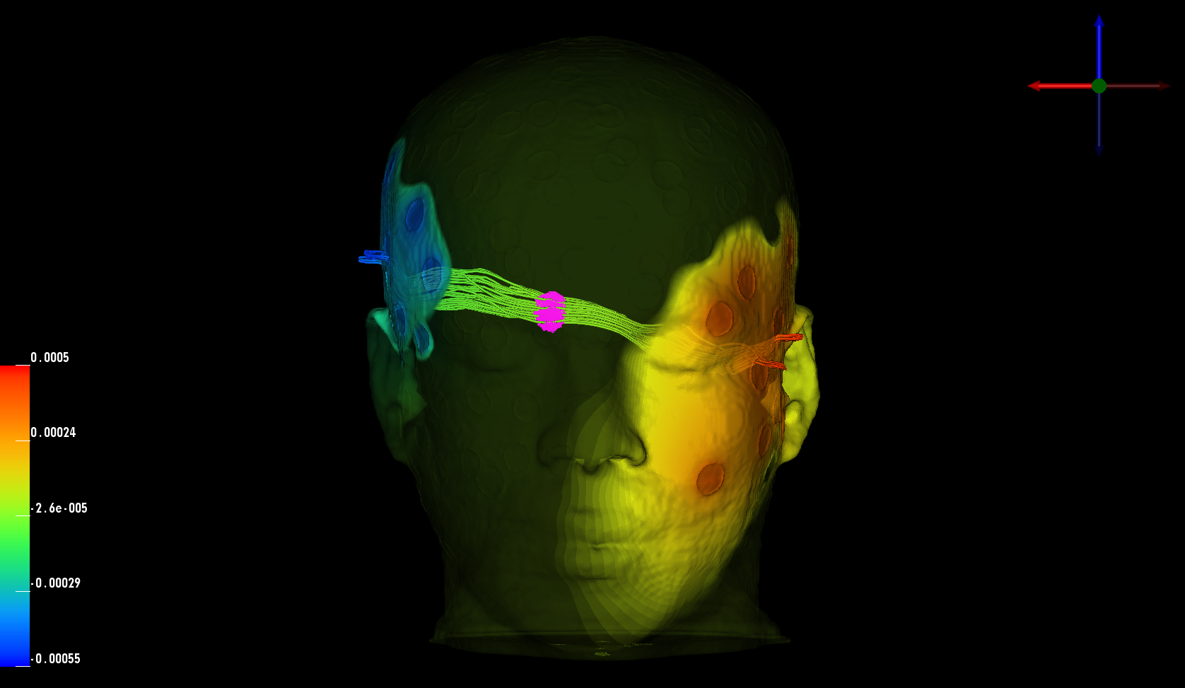 In collaboration with Dr. Don Tucker and his colleagues at Electrical Geodesics Inc (EGI) and the University of Oregon, this DBP is concerned with improving our ability to reconstruct and visualize neuroelectric sources (source localization) from EEG measurements and also our ability to stimulate specific brain regions using electrodes attached on to the scalp of the subject (transcranial direct current stimulation, tDCS).
For both research and clinical practice, EEG is a cost-effective tool to understand and excite brain activity. EEG advances have significantly improved the spatial resolution of source estimates and offer the promise of precise spatio-temporal monitoring and stimulation of cortical brain activity. By itself, high-resolution EEG would be affordable even for small hospitals in remote locations and could be easily managed by technicians in the field.
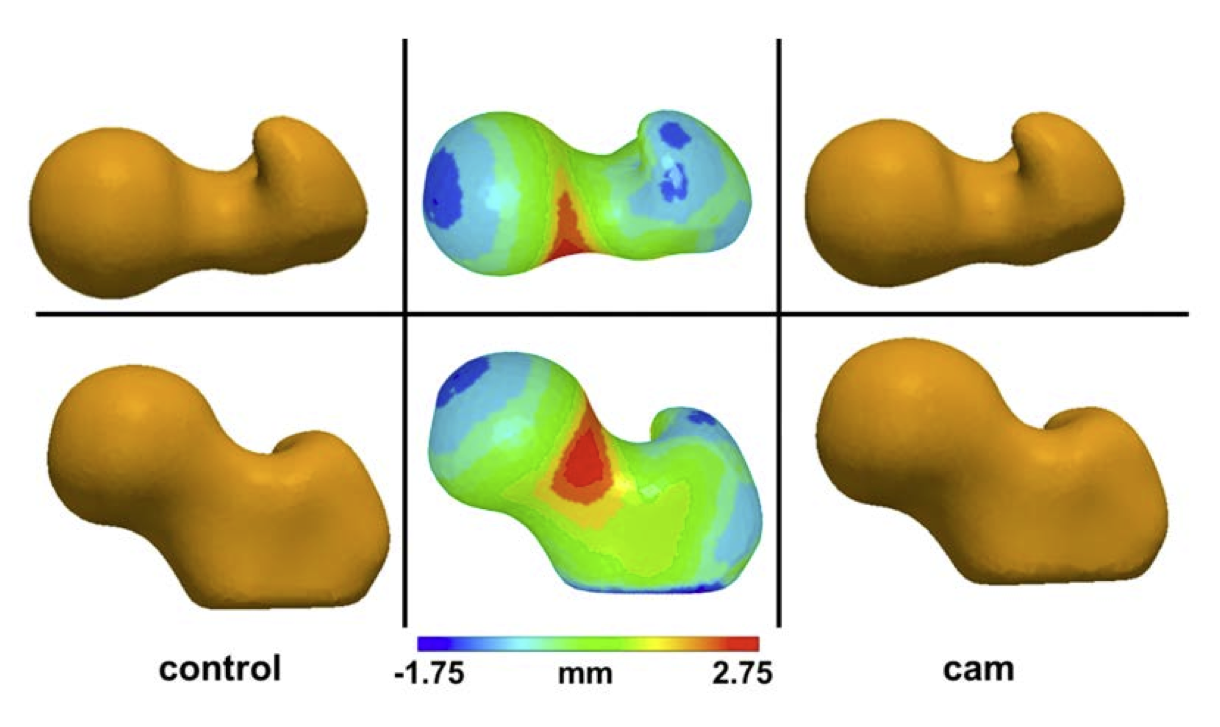
Cam Femoroacetabular Impingement Analysis using Statistical Shape Modeling
 |
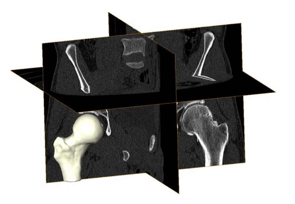 |
| Mean control (left) and cam (right) shapes. Middle images show the mean control shape with color plots depicting how the mean cam shape differed across the femoral head, neck, and proximal shaft. Top and bottom rows show different rotations of the femoral head. | Volumetric CT images from a cam FAI patient. Validated threshold settings were applied to CT images to segment and reconstruct the bony morphology of each femur. |