


SCI Publications
2014


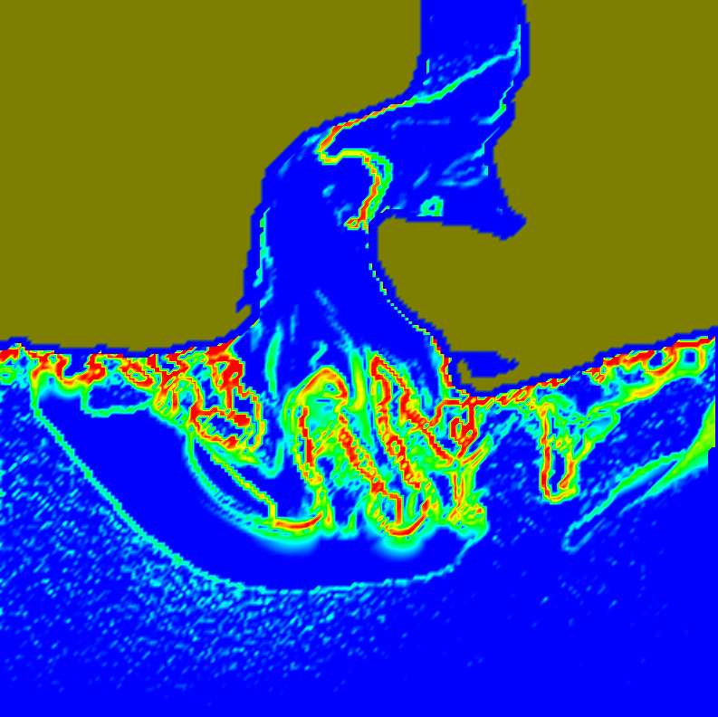
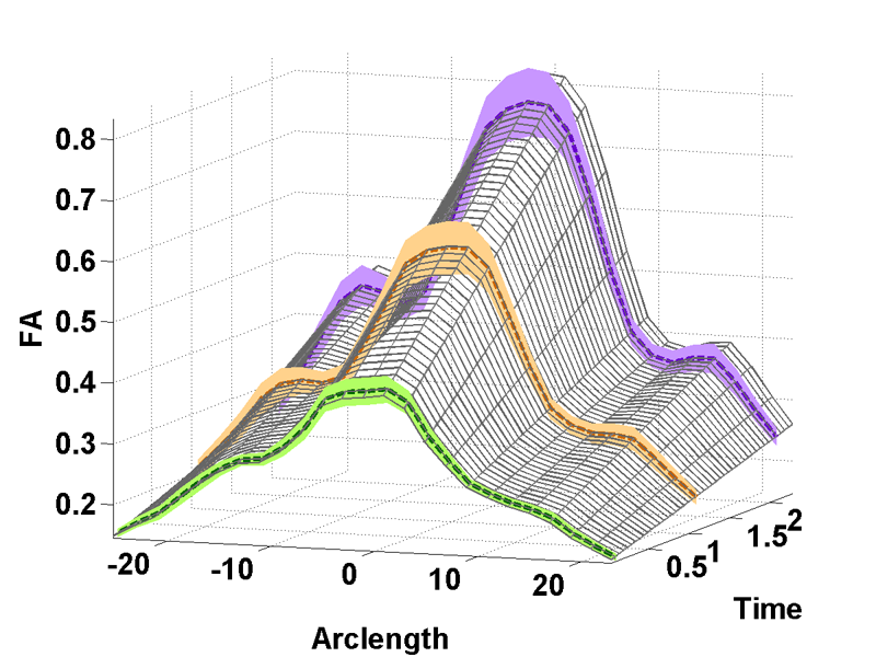
M. Mirzargar, R. Whitaker, R. M. Kirby.
“Curve Boxplot: Generalization of Boxplot for Ensembles of Curves,” In IEEE Transactions on Visualization and Computer Graphics, Vol. 20, No. 12, IEEE, pp. 2654-63. December, 2014.
In simulation science, computational scientists often study the behavior of their simulations by repeated solutions with variations in parameters and/or boundary values or initial conditions. Through such simulation ensembles, one can try to understand or quantify the variability or uncertainty in a solution as a function of the various inputs or model assumptions. In response to a growing interest in simulation ensembles, the visualization community has developed a suite of methods for allowing users to observe and understand the properties of these ensembles in an efficient and effective manner. An important aspect of visualizing simulations is the analysis of derived features, often represented as points, surfaces, or curves. In this paper, we present a novel, nonparametric method for summarizing ensembles of 2D and 3D curves. We propose an extension of a method from descriptive statistics, data depth, to curves. We also demonstrate a set of rendering and visualization strategies for showing rank statistics of an ensemble of curves, which is a generalization of traditional whisker plots or boxplots to multidimensional curves. Results are presented for applications in neuroimaging, hurricane forecasting and fluid dynamics
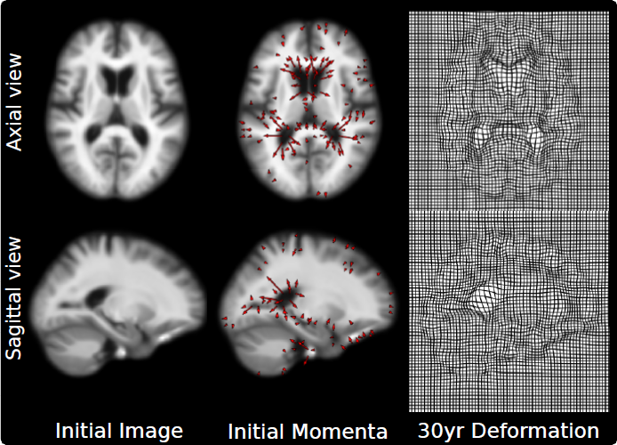
P. Muralidharan, J. Fishbaugh, H.J. Johnson, S. Durrleman, J.S. Paulsen, G. Gerig, P.T. Fletcher.
“Diffeomorphic Shape Trajectories for Improved Longitudinal Segmentation and Statistics,” In Proceedings of Medical Image Computing and Computer Assisted Intervention (MICCAI), 2014.

J.A. Nielsen, B.A. Zielinski, P.T. Fletcher, A.L. Alexander, N. Lange, E.D. Bigler, J.E. Lainhart, J.S. Anderson.
“Abnormal lateralization of functional connectivity between language and default mode regions in autism,” In Molecular Autism, Vol. 5, No. 1, pp. 8. 2014.
DOI: 10.1186/2040-2392-5-8
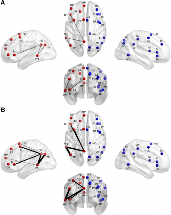
Methods: Using functional connectivity magnetic resonance imaging from a large publicly available sample (n = 964), we tested whether abnormal functional lateralization in autism exists preferentially in language regions or in a more diffuse pattern across networks of lateralized brain regions.
Results: The autism group exhibited significantly reduced left lateralization in a few connections involving language regions and regions from the default mode network, but results were not significant throughout left- and right-lateralized networks. There is a trend that suggests the lack of left lateralization in a connection involving Wernicke area and the posterior cingulate cortex associates with more severe autism.
Conclusions: Abnormal language lateralization in autism may be due to abnormal language development rather than to a deficit in hemispheric specialization of the entire brain.
Keywords: brain lateralization, brain asymmetry, autism, autism spectrum disorder, language, functional magnetic resonance imaging, functional connectivity
K.A. Nestor, J.D. Jones, C.R. Butson, T. Morishita, C.E. Jacobson, D.A. Peace, D. Chen, K.D. Foote, M.S. Okun.
“Coordinate-based lead location does not predict Parkinson's disease deep brain stimulation outcome,” In PloS One, Vol. 9, No. 4, pp. e93524. January, 2014.
ISSN: 1932-6203
DOI: 10.1371/journal.pone.0093524
PubMed ID: 24691109
BACKGROUND: Effective target regions for deep brain stimulation (DBS) in Parkinson's disease (PD) have been well characterized. We sought to study whether the measured Cartesian coordinates of an implanted DBS lead are predictive of motor outcome(s). We tested the hypothesis that the position and trajectory of the DBS lead relative to the mid-commissural point (MCP) are significant predictors of clinical outcomes. We expected that due to neuroanatomical variation among individuals, a simple measure of the position of the DBS lead relative to MCP (commonly used in clinical practice) may not be a reliable predictor of clinical outcomes when utilized alone.
METHODS: 55 PD subjects implanted with subthalamic nucleus (STN) DBS and 41 subjects implanted with globus pallidus internus (GPi) DBS were included. Lead locations in AC-PC space (x, y, z coordinates of the active contact and sagittal and coronal entry angles) measured on high-resolution CT-MRI fused images, and motor outcomes (Unified Parkinson's Disease Rating Scale) were analyzed to confirm or refute a correlation between coordinate-based lead locations and DBS motor outcomes.
RESULTS: Coordinate-based lead locations were not a significant predictor of change in UPDRS III motor scores when comparing pre- versus post-operative values. The only potentially significant individual predictor of change in UPDRS motor scores was the antero-posterior coordinate of the GPi lead (more anterior lead locations resulted in a worse outcome), but this was only a statistical trend (p<.082).
CONCLUSION: The results of the study showed that a simple measure of the position of the DBS lead relative to the MCP is not significantly correlated with PD motor outcomes, presumably because this method fails to account for individual neuroanatomical variability. However, there is broad agreement that motor outcomes depend strongly on lead location. The results suggest the need for more detailed identification of stimulation location relative to anatomical targets.
I. Oguz, M. Farzinfar, J. Matsui, F. Budin, Z. Liu, G. Gerig, H.J. Johnson, M.A. Styner.
“DTIPrep: Quality Control of Diffusion-Weighted Images,” In Frontiers in Neuroinformatics, Vol. 8, No. 4, 2014.
DOI: 10.3389/fninf.2014.00004
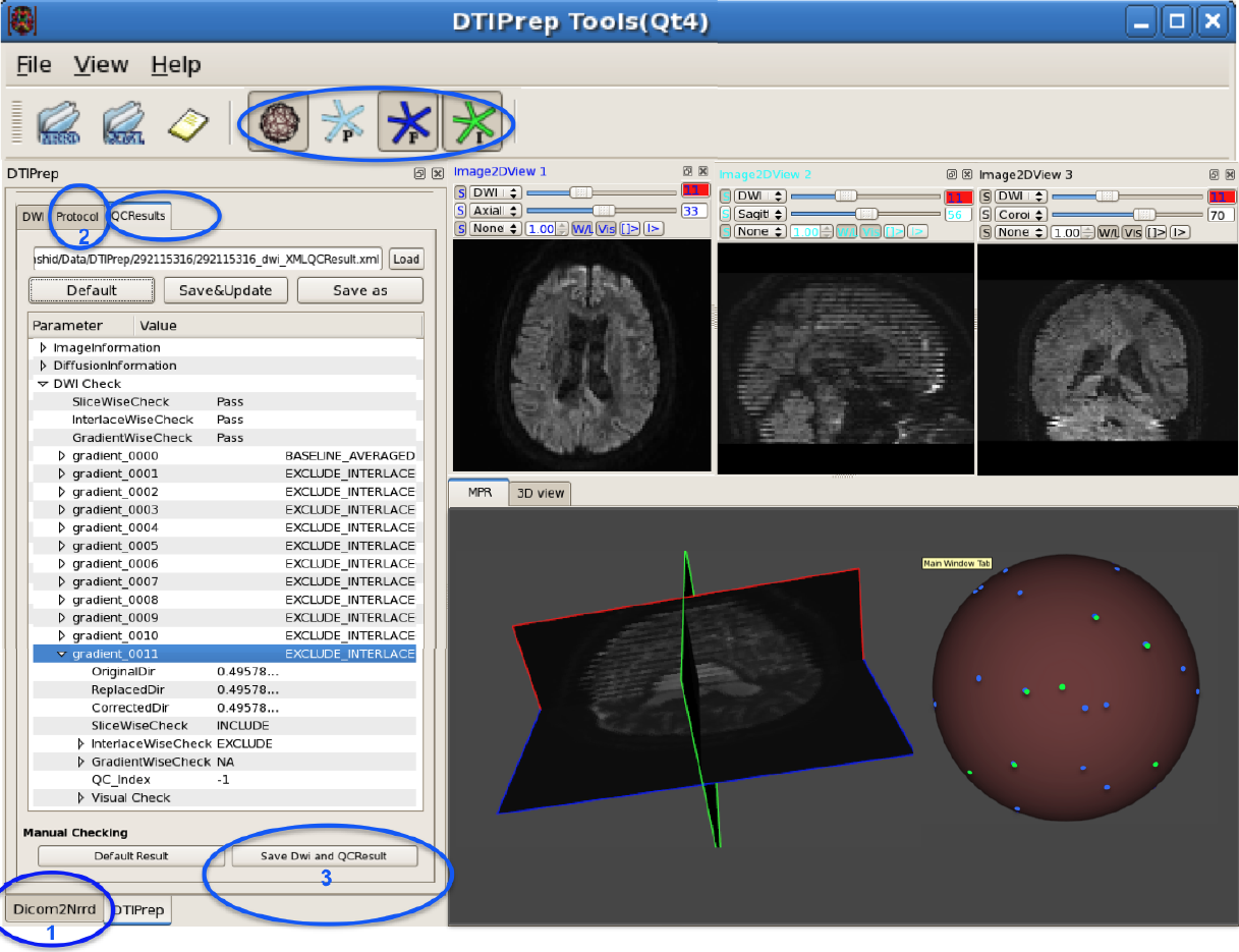
In the last decade, diffusion MRI (dMRI) studies of the human and animal brain have been used to investigate a multitude of pathologies and drug-related effects in neuroscience research. Study after study identifies white matter (WM) degeneration as a crucial biomarker for all these diseases. The tool of choice for studying WM is dMRI. However, dMRI has inherently low signal-to-noise ratio and its acquisition requires a relatively long scan time; in fact, the high loads required occasionally stress scanner hardware past the point of physical failure. As a result, many types of artifacts implicate the quality of diffusion imagery. Using these complex scans containing artifacts without quality control (QC) can result in considerable error and bias in the subsequent analysis, negatively affecting the results of research studies using them. However, dMRI QC remains an under-recognized issue in the dMRI community as there are no user-friendly tools commonly available to comprehensively address the issue of dMRI QC. As a result, current dMRI studies often perform a poor job at dMRI QC.
Thorough QC of diffusion MRI will reduce measurement noise and improve reproducibility, and sensitivity in neuroimaging studies; this will allow researchers to more fully exploit the power of the dMRI technique and will ultimately advance neuroscience. Therefore, in this manuscript, we present our open-source software, DTIPrep, as a unified, user friendly platform for thorough quality control of dMRI data. These include artifacts caused by eddy-currents, head motion, bed vibration and pulsation, venetian blind artifacts, as well as slice-wise and gradient-wise intensity inconsistencies. This paper summarizes a basic set of features of DTIPrep described earlier and focuses on newly added capabilities related to directional artifacts and bias analysis.
Keywords: diffusion MRI, Diffusion Tensor Imaging, Quality control, Software, open-source, preprocessing
D.C.B. de Oliveira, A. Humphrey, Q. Meng, Z. Rakamaric, M. Berzins, G. Gopalakrishnan.
“Systematic Debugging of Concurrent Systems Using Coalesced Stack Trace Graphs,” In Proceedings of the 27th International Workshop on Languages and Compilers for Parallel Computing (LCPC), September, 2014.
A central need during software development of large-scale parallel systems is tools that help help to identify the root causes of bugs quickly. Given the massive scale of these systems, tools that highlight changes--say introduced across software versions or their operating conditions (e.g., inputs, schedules)--can prove to be highly effective in practice. Conventional debuggers, while good at presenting details at the problem-site (e.g., crash), often omit contextual information to identify the root causes of the bug. We present a new approach to collect and coalesce stack traces, leading to an efficient summary display of salient system control flow differences in a graphical form called Coalesced Stack Trace Graphs (CSTG). CSTGs have helped us understand and debug situations within a computational framework called Uintah that has been deployed at large scale, and undergoes frequent version updates. In this paper, we detail CSTGs through case studies in the context of Uintah where unexpected behaviors caused by different vesions of software or occurring across different time-steps of a system (e.g., due to non-determinism) are debugged. We show that CSTG also gives conventional debuggers a far more productive and guided role to play.
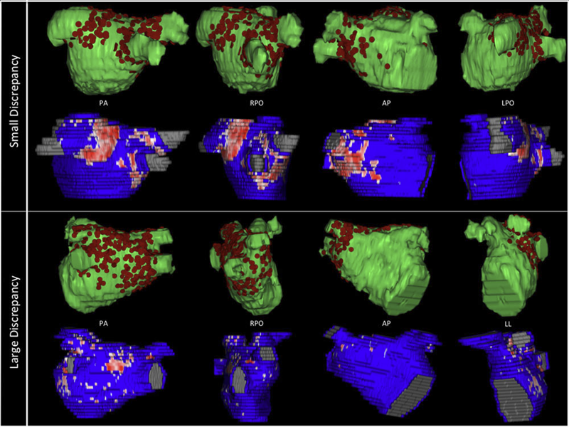
B.R. Parmar, T.R. Jarrett, N.S. Burgon, E.G. Kholmovski, N.W. Akoum, N. Hu, R.S. Macleod, N.F. Marrouche, R. Ranjan.
“Comparison of Left Atrial Area Marked Ablated in Electroanatomical Maps with Scar in MRI,” In Journal of Cardiovascular Electrophysiology, 2014.
DOI: 10.1111/jce.12357
Background
Three-dimensional electroanatomic mapping (EAM) is routinely used to mark ablated areas during radiofrequency ablation. We hypothesized that, in atrial fibrillation (AF) ablation, EAM overestimates scar formation in the left atrium (LA) when compared to the scar seen on late-gadolinium enhancement magnetic resonance imaging (LGE-MRI).
Methods and Results
Of the 235 patients who underwent initial ablation for AF at our institution between August 2011 and December 2012, we retrospectively identified 70 patients who had preprocedural magnetic resonance angiography merged with LA anatomy in EAM software and had a 3-month postablation LGE-MRI for assessment of scar. Ablated area was marked intraprocedurally using EAM software and quantified retrospectively. Scarred area was quantified in 3-month postablation LGE-MRI. The mean ablated area in EAM was 30.5 ± 7.5% of the LA endocardial surface and the mean scarred area in LGE-MRI was 13.9 ± 5.9% (P < 0.001). This significant difference in the ablated area marked in the EAM and scar area in the LGE-MRI was present for each of the 3 independent operators. Complete pulmonary vein (PV) encirclement representing electrical isolation was observed in 87.8% of the PVs in EAM as compared to only 37.4% in LGE-MRI (P < 0.001).
Conclusions
In AF ablation, EAM significantly overestimates the resultant scar as assessed with a follow-up LGE-MRI.
Keywords: atrial fibrillation, magnetic resonance imaging, radiofrequency ablation
Christian Partl, Alexander Lex, Marc Streit, Hendrik Strobelt, Anne-Mai Wasserman, Hanspeter Pfister,, Dieter Schmalstieg.
“ConTour: Data-Driven Exploration of Multi-Relational Datasets for Drug Discovery,” In IEEE Transactions on Visualization and Computer Graphics (VAST '14), Vol. 20, No. 12, pp. 1883--1892. 2014.
ISSN: 1077-2626
DOI: 10.1109/TVCG.2014.2346752
Large scale data analysis is nowadays a crucial part of drug discovery. Biologists and chemists need to quickly explore and evaluate potentially effective yet safe compounds based on many datasets that are in relationship with each other. However, there is a lack of tools that support them in these processes. To remedy this, we developed ConTour, an interactive visual analytics technique that enables the exploration of these complex, multi-relational datasets. At its core ConTour lists all items of each dataset in a column. Relationships between the columns are revealed through interaction: selecting one or multiple items in one column highlights and re-sorts the items in other columns. Filters based on relationships enable drilling down into the large data space. To identify interesting items in the first place, ConTour employs advanced sorting strategies, including strategies based on connectivity strength and uniqueness, as well as sorting based on item attributes. ConTour also introduces interactive nesting of columns, a powerful method to show the related items of a child column for each item in the parent column. Within the columns, ConTour shows rich attribute data about the items as well as information about the connection strengths to other datasets. Finally, ConTour provides a number of detail views, which can show items from multiple datasets and their associated data at the same time. We demonstrate the utility of our system in case studies conducted with a team of chemical biologists, who investigate the effects of chemical compounds on cells and need to understand the underlying mechanisms.
A. J. Perez, M. Seyedhosseini, T. J. Deerinck, E. A. Bushong, S. Panda, T. Tasdizen, M. H. Ellisman.
“A workflow for the automatic segmentation of organelles in electron microscopy image stacks,” In Frontiers in Neuroanatomy, Vol. 8, No. 126, 2014.
DOI: 10.3389/fnana.2014.00126
Electron microscopy (EM) facilitates analysis of the form, distribution, and functional status of key organelle systems in various pathological processes, including those associated with neurodegenerative disease. Such EM data often provide important new insights into the underlying disease mechanisms. The development of more accurate and efficient methods to quantify changes in subcellular microanatomy has already proven key to understanding the pathogenesis of Parkinson's and Alzheimer's diseases, as well as glaucoma. While our ability to acquire large volumes of 3D EM data is progressing rapidly, more advanced analysis tools are needed to assist in measuring precise three-dimensional morphologies of organelles within data sets that can include hundreds to thousands of whole cells. Although new imaging instrument throughputs can exceed teravoxels of data per day, image segmentation and analysis remain significant bottlenecks to achieving quantitative descriptions of whole cell structural organellomes. Here, we present a novel method for the automatic segmentation of organelles in 3D EM image stacks. Segmentations are generated using only 2D image information, making the method suitable for anisotropic imaging techniques such as serial block-face scanning electron microscopy (SBEM). Additionally, no assumptions about 3D organelle morphology are made, ensuring the method can be easily expanded to any number of structurally and functionally diverse organelles. Following the presentation of our algorithm, we validate its performance by assessing the segmentation accuracy of different organelle targets in an example SBEM dataset and demonstrate that it can be efficiently parallelized on supercomputing resources, resulting in a dramatic reduction in runtime.
A. Perez, M. Seyedhosseini, T. Tasdizen, M. Ellisman.
“Automated workflows for the morphological characterization of organelles in electron microscopy image stacks (LB72),” In The FASEB Journal, Vol. 28, No. 1 Supplement LB72, April, 2014.
Advances in three-dimensional electron microscopy (EM) have facilitated the collection of image stacks with a field-of-view that is large enough to cover a significant percentage of anatomical subdivisions at nano-resolution. When coupled with enhanced staining protocols, such techniques produce data that can be mined to establish the morphologies of all organelles across hundreds of whole cells in their in situ environments. Although instrument throughputs are approaching terabytes of data per day, image segmentation and analysis remain significant bottlenecks in achieving quantitative descriptions of whole cell organellomes. Here we describe computational workflows that achieve the automatic segmentation of organelles from regions of the central nervous system by applying supervised machine learning algorithms to slices of serial block-face scanning EM (SBEM) datasets. We also demonstrate that our workflows can be parallelized on supercomputing resources, resulting in a dramatic reduction of their run times. These methods significantly expedite the development of anatomical models at the subcellular scale and facilitate the study of how these models may be perturbed following pathological insults.
N. Ramesh, T. Tasdizen.
“Cell tracking using particle filters with implicit convex shape model in 4D confocal microscopy images,” In 2014 IEEE International Conference on Image Processing (ICIP), IEEE, Oct, 2014.
DOI: 10.1109/icip.2014.7025089
Bayesian frameworks are commonly used in tracking algorithms. An important example is the particle filter, where a stochastic motion model describes the evolution of the state, and the observation model relates the noisy measurements to the state. Particle filters have been used to track the lineage of cells. Propagating the shape model of the cell through the particle filter is beneficial for tracking. We approximate arbitrary shapes of cells with a novel implicit convex function. The importance sampling step of the particle filter is defined using the cost associated with fitting our implicit convex shape model to the observations. Our technique is capable of tracking the lineage of cells for nonmitotic stages. We validate our algorithm by tracking the lineage of retinal and lens cells in zebrafish embryos.
F. Rousset, C. Vachet, C. Conlin, M. Heilbrun, J.L. Zhang, V.S. Lee, G. Gerig.
“Semi-automated application for kidney motion correction and filtration analysis in MR renography,” In Proceeding of the 2014 Joint Annual Meeting ISMRM-ESMRMB, pp. (accepted). 2014.
Altered renal function commonly affects patients with cirrhosis, a consequence of chronic liver disease. From lowdose contrast material-enhanced magnetic resonance (MR) renography, we can estimate the Glomerular Filtration Rate (GFR), an important parameter to assess renal function. Two-dimensional MR images are acquired every 2 seconds for approximately 5 minutes during free breathing, which results in a dynamic series of 140 images representing kidney filtration over time. This specific acquisition presents dynamic contrast changes but is also challenged by organ motion due to breathing. Rather than use conventional image registration techniques, we opted for an alternative method based on object detection. We developed a novel analysis framework available under a stand-alone toolkit to efficiently register dynamic kidney series, manually select regions of interest, visualize the concentration curves for these ROIs, and fit them into a model to obtain GFR values. This open-source cross-platform application is written in C++, using the Insight Segmentation and Registration Toolkit (ITK) library, and QT4 as a graphical user interface.
N. Sadeghi, J.H. Gilmore, W. Lin, G. Gerig.
“Normative Modeling of Early Brain Maturation from Longitudinal DTI Reveals Twin-Singleton Differences,” In Proceeding of the 2014 Joint Annual Meeting ISMRM-ESMRMB, pp. (accepted). 2014.
Early brain development of white matter is characterized by rapid organization and structuring. Magnetic Resonance diffusion tensor imaging (MR-DTI) provides the possibility of capturing these changes non-invasively by following individuals longitudinally in order to better understand departures from normal brain development in subjects at risk for mental illness [1]. Longitudinal imaging of individuals suggests the use of 4D (3D, time) image analysis and longitudinal statistical modeling [3].
N. Sadeghi, P.T. Fletcher, M. Prastawa, J.H. Gilmore, G. Gerig.
“Subject-specific prediction using nonlinear population modeling: Application to early brain maturation from DTI,” In Proceedings of Medical Image Computing and Computer-Assisted Intervention (MICCAI 2014), 2014.
A.R. Sanderson.
“An Alternative Formulation of Lyapunov Exponents for Computing Lagrangian Coherent Structures,” In Proceedings of the 2014 IEEE Pacific Visualization Symposium (PacificVis), Yokahama Japan, 2014.
M. Seyedhosseini, T. Tasdizen.
“Disjunctive normal random forests,” In Pattern Recognition, September, 2014.
DOI: 10.1016/j.patcog.2014.08.023
We develop a novel supervised learning/classification method, called disjunctive normal random forest (DNRF). A DNRF is an ensemble of randomly trained disjunctive normal decision trees (DNDT). To construct a DNDT, we formulate each decision tree in the random forest as a disjunction of rules, which are conjunctions of Boolean functions. We then approximate this disjunction of conjunctions with a differentiable function and approach the learning process as a risk minimization problem that incorporates the classification error into a single global objective function. The minimization problem is solved using gradient descent. DNRFs are able to learn complex decision boundaries and achieve low generalization error. We present experimental results demonstrating the improved performance of DNDTs and DNRFs over conventional decision trees and random forests. We also show the superior performance of DNRFs over state-of-the-art classification methods on benchmark datasets.
Keywords: Random forest, Decision tree, Classifier, Supervised learning, Disjunctive normal form
M. Seyedhosseini, T. Tasdizen.
“Scene Labeling with Contextual Hierarchical Models,” In CoRR, Vol. abs/1402.0595, 2014.

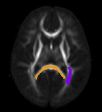
A. Sharma, P.T. Fletcher, J.H. Gilmore, M.L. Escolar, A. Gupta, M. Styner, G. Gerig.
“Parametric Regression Scheme for Distributions: Analysis of DTI Fiber Tract Diffusion Changes in Early Brain Development,” In Proceedings of the 2014 IEEE International Symposium on Biomedical Imaging (ISBI), pp. (accepted). 2014.
Keywords: linear regression, distribution-valued data, spatiotemporal growth trajectory, DTI, early neurodevelopment
R. Sicat, J. Krüger, T. Möller, M. Hadwiger.
“Sparse PDF Volumes for Consistent Multi-Resolution Volume Rendering,” In IEEE Transactions on Visualization and Computer Graphics (TVCG), Vol. PP, No. 99, pp. 1--1. 2014.
ISBN: 1077-2626
DOI: 10.1109/TVCG.2014.2346324
This paper presents a new multi-resolution volume representation called sparse pdf volumes, which enables consistent multi-resolution volume rendering based on probability density functions (pdfs) of voxel neighborhoods. These pdfs are defined in the 4D domain jointly comprising the 3D volume and its 1D intensity range. Crucially, the computation of sparse pdf volumes exploits data coherence in 4D, resulting in a sparse representation with surprisingly low storage requirements. At run time, we dynamically apply transfer functions to the pdfs using simple and fast convolutions. Whereas standard low-pass filtering and down-sampling incur visible differences between resolution levels, the use of pdfs facilitates consistent results independent of the resolution level used. We describe the efficient out-of-core computation of large-scale sparse pdf volumes, using a novel iterative simplification procedure of a mixture of 4D Gaussians. Finally, our data structure is optimized to facilitate interactive multi-resolution volume rendering on GPUs.
N.P. Singh, J. Hinkle, S. Joshi, P.T. Fletcher.
“An Efficient Parallel Algorithm for Hierarchical Geodesic Models in Diffeomorphisms,” In Proceedings of the 2014 IEEE International Symposium on Biomedical Imaging (ISBI), pp. (accepted). 2014.
Keywords: LDDMM, HGM, Vector Momentum, Diffeomorphisms, Longitudinal Analysis