SCI Publications
2014


P. Skraba, Bei Wang, G. Chen, P. Rosen.
“2D Vector Field Simplification Based on Robustness,” In Proceedings of the 2014 IEEE Pacific Visualization Symposium, PacificVis, Note: Awarded Best Paper!, 2014.
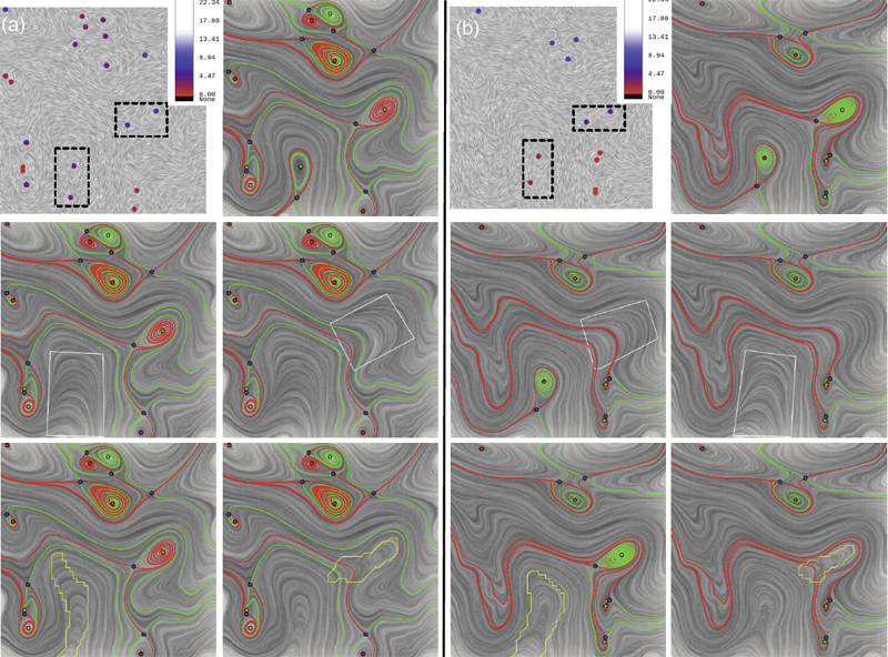
Keywords: vector field, topology-based techniques, flow visualization
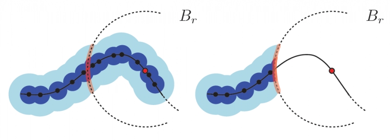
P. Skraba, Bei Wang.
“Interpreting Feature Tracking Through the Lens of Robustness,” In Mathematics and Visualization, Springer, pp. 19-37. 2014.
DOI: 10.1007/978-3-319-04099-8_2
P. Skraba, Bei Wang.
“Approximating Local Homology from Samples,” In Proceedings 25th Annual ACM-SIAM Symposium on Discrete Algorithms (SODA), pp. 174-192. 2014.
J. Sourati, D. Erdogmus, J.G. Dy, D.H. Brooks.
“Accelerated learning-based interactive image segmentation using pairwise constraints,” In IEEE Transactions on Medical Image Processing, Vol. 23, No. 7, pp. 3057-3070. July, 2014.
DOI: 10.1109/TIP.2014.2325783
PubMed ID: 24860031
PubMed Central ID: PMC4096329
Algorithms for fully automatic segmentation of images are often not sufficiently generic with suitable accuracy, and fully manual segmentation is not practical in many settings. There is a need for semiautomatic algorithms, which are capable of interacting with the user and taking into account the collected feedback. Typically, such methods have simply incorporated user feedback directly. Here, we employ active learning of optimal queries to guide user interaction. Our work in this paper is based on constrained spectral clustering that iteratively incorporates user feedback by propagating it through the calculated affinities. The original framework does not scale well to large data sets, and hence is not straightforward to apply to interactive image segmentation. In order to address this issue, we adopt advanced numerical methods for eigen-decomposition implemented over a subsampling scheme. Our key innovation, however, is an active learning strategy that chooses pairwise queries to present to the user in order to increase the rate of learning from the feedback. Performance evaluation is carried out on the Berkeley segmentation and Graz-02 image data sets, confirming that convergence to high accuracy levels is realizable in relatively few iterations.
R. Stoll, E. Pardyjak, J.J. Kim, T. Harman, A.N. Hayati.
“An inter-model comparison of three computation fluid dynamics techniques for step-up and step-down street canyon flows,” In ASME FEDSM/ICNMM symposium on urban fluid mechanics, August, 2014.
M. Streit, A. Lex, S. Gratzl, C. Partl, D. Schmalstieg, H. Pfister, P. J. Park,, N. Gehlenborg.
“Guided visual exploration of genomic stratifications in cancer,” In Nature Methods, Vol. 11, No. 9, pp. 884--885. Sep, 2014.
ISSN: 1548-7091
DOI: 10.1038/nmeth.3088
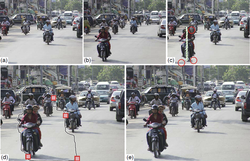
B. Summa, A.A. Gooch, G. Scorzelli, V. Pascucci.
“Towards Paint and Click: Unified Interactions for Image Boundaries,” SCI Technical Report, No. UUSCI-2014-004, SCI Institute, University of Utah, December, 2014.
T. Tasdizen, M. Seyedhosseini, T. Liu, C. Jones, E. Jurrus.
“Image Segmentation for Connectomics Using Machine Learning,” In Computational Intelligence in Biomedical Imaging, Edited by Suzuki, Kenji, Springer New York, pp. 237--278. 2014.
ISBN: 978-1-4614-7244-5
DOI: 10.1007/978-1-4614-7245-2_10
Reconstruction of neural circuits at the microscopic scale of individual neurons and synapses, also known as connectomics, is an important challenge for neuroscience. While an important motivation of connectomics is providing anatomical ground truth for neural circuit models, the ability to decipher neural wiring maps at the individual cell level is also important in studies of many neurodegenerative diseases. Reconstruction of a neural circuit at the individual neuron level requires the use of electron microscopy images due to their extremely high resolution. Computational challenges include pixel-by-pixel annotation of these images into classes such as cell membrane, mitochondria and synaptic vesicles and the segmentation of individual neurons. State-of-the-art image analysis solutions are still far from the accuracy and robustness of human vision and biologists are still limited to studying small neural circuits using mostly manual analysis. In this chapter, we describe our image analysis pipeline that makes use of novel supervised machine learning techniques to tackle this problem.
C. Turkay, A. Lex, M. Streit, H. Pfister,, H. Hauser.
“Characterizing Cancer Subtypes using Dual Analysis in Caleydo,” In IEEE Computer Graphics and Applications, Vol. 34, No. 2, pp. 38--47. March, 2014.
ISSN: 0272-1716
DOI: 10.1109/MCG.2014.1
Dual analysis uses statistics to describe both the dimensions and rows of a high-dimensional dataset. Researchers have integrated it into StratomeX, a Caleydo view for cancer subtype analysis. In addition, significant-difference plots show the elements of a candidate subtype that differ significantly from other subtypes, thus letting analysts characterize subtypes. Analysts can also investigate how data samples relate to their assigned subtype and other groups. This approach lets them create well-defined subtypes based on statistical properties. Three case studies demonstrate the approach's utility, showing how it reproduced findings from a published subtype characterization.
C.J. Underwood, L.T. Edgar, J.B. Hoying, J.A. Weiss.
“Cell-generated traction forces and the resulting matrix deformation modulate microvascular alignment and growth during angiogenesis,” In American Journal of Physiology: Heart and Circulatory Physiology, Vol. 307, No. H152-H164, 2014.
DOI: 10.1152/ajpheart.00995.2013
PubMed ID: 24816262
PubMed Central ID: PMC4101638
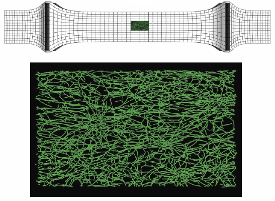
Keywords: angiogenesis, deformation, image analysis, morphometry, orientation, strain
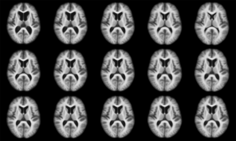
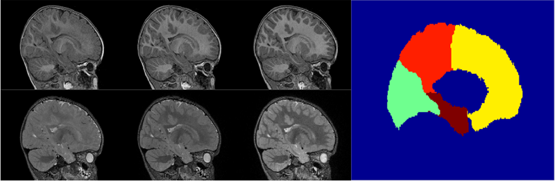
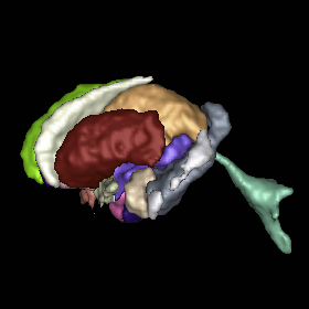
C. Vachet, H.C. Hazlett, J. Piven, G. Gerig.
“4D Modeling of Infant Brain Growth in Down's Syndrome and Controls from longitudinal MRI,” In Proceeding of the 2014 Joint Annual Meeting ISMRM-ESMRMB, pp. (accepted). 2014.
Modeling of early brain growth trajectories from longitudinal MRI will provide new insight into neurodevelopmental characteristics, timing and type of changes in neurological disorders from controls. In addition to an ongoing large-scale infant autism neuroimaging study 1, we recruited 4 infants with Down’s syndrome (DS) in order to evaluate newly developed methods for 4D segmentation from longitudinal infant MRI, and for temporal modeling of brain growth trajectories. Specifically to Down's, a comparison of patterns of full brain and lobar tissue growth may lead to better insight into the observed variability of cognitive development and neurological effects, and may help with development of disease-modifying therapeutic intervention.
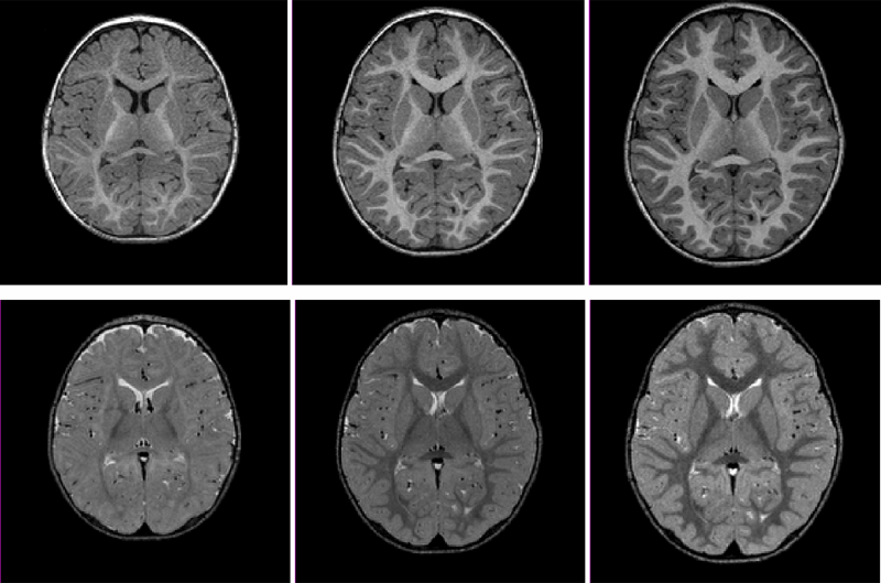
A. Vardhan, M. Prastawa, C. Vachet, J. Piven, G. Gerig.
“Characterizing growth patterns in longitudinal MRI using image contrast,” In Proceedings of Medical Imaging 2014: Image Processing, 2014.
A. Vardhan, N. Sadeghi, C. Vachet, J. Piven, G. Gerig.
“Joint Longitudinal Modeling of Brain Appearance in Multimodal MRI for the Characterization of Early Brain Developmental Processes,” In Spatiotemporal Image Analysis for Longitudinal and Time-Series Image Data (STIA'14) , LNCS. MICCAI'14, Springer Verlag, June, 2014.
A.R. Verde, F. Budin, J.-B. Berger, A. Gupta, M. Farzinfar, A. Kaiser, M. Ahn, H. Johnson, J. Matsui, H.C. Hazlett, A. Sharma, C. Goodlett, Y. Shi, S. Gouttard, C. Vachet, J. Piven, H. Zhu, G. Gerig, M. Styner.
“UNC-Utah NA-MIC framework for DTI fiber tract analysis,” In Frontiers in Neuroinformatics, Vol. 7, No. 51, January, 2014.
DOI: 10.3389/fninf.2013.00051
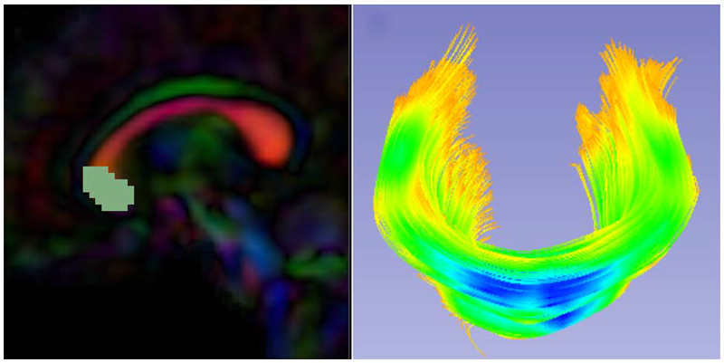
Keywords: neonatal neuroimaging, white matter pathways, magnetic resonance imaging, diffusion tensor imaging, diffusion imaging quality control, DTI atlas building
Bo Wang, W. Liu, M. Prastawa, A. Irimia, P.M. Vespa, J.D. van Horn, P.T. Fletcher, G. Gerig.
“4D Active Cut: An Interactive Tool for Pathological Anatomy Modeling,” In Proceedings of the 2014 IEEE International Symposium on Biomedical Imaging (ISBI), pp. (accepted). 2014.

Keywords: Active learning, graph cuts, longitudinal MRI, Markov Random Fields, semi-supervised learning
J. Wang, C. Vachet, A. Rumple, S. Gouttard, C. Ouzie, E. Perrot, G. Du, X. Huang, G. Gerig, M.A. Styner.
“Multi-atlas segmentation of subcortical brain structures via the AutoSeg software pipeline,” In Frontiers in Neuroinformatics, Vol. 8, No. 7, 2014.
DOI: 10.3389/fninf.2014.00007
Keywords: segmentation, Registration, MRI, Atlas, Brain, Insight Toolkit
Y. Wan, H. Otsuna, K. Kwan, C.D. Hansen.
“Real-Time Dense Nucleus Selection from Confocal Data,” In Proceedings of the Eurographics Workshop on Visual Computing for Biology and Medicine, 2014.
Selecting structures from volume data using direct over-the-visualization interactions, such as a paint brush, is perhaps the most intuitive method in a variety of application scenarios. Unfortunately, it seems difficult to design a universal tool that is effective for all different structures in biology research. In [WOCH12b], an interactive technique was proposed for extracting neural structures from confocal microscopy data. It uses a dual-stroke paint brush to select desired structures directly from volume visualizations. However, the technique breaks down when it was applied to selecting densely packed structures with condensed shapes, such as nuclei from zebrafish eye development research. We collaborated with biologists studying zebrafish eye development and adapted the paint brush tool for real-time nucleus selection from volume data. The morphological diffusion algorithm used in the previous paint brush is restricted to gradient descending directions for improved nucleus boundary definition. Occluded seeds are removed using backward ray-casting. The adapted paint brush is then used in tracking cell movements in a time sequence dataset of a developing zebrafish eye.
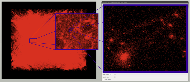
W. Widanagamaachchi, P.-T. Bremer, C. Sewell, L.-T. Lo; J. Ahrens, V. Pascucci.
“Data-Parallel Halo Finding with Variable Linking Lengths,” In Proceedings of the 2014 IEEE 4th Symposium on Large Data Analysis and Visualization (LDAV), pp. 27--34. November, 2014.
Y.-Y. Yu, P.T. Fletcher, S.P. Awate.
“Hierarchical Bayesian Modeling, Estimation, and Sampling for Multigroup Shape Analysis,” In Proceedings of Medical Image Computing and Computer Assisted Intervention (MICCAI), 2014.
This paper proposes a novel method for the analysis of anatomical shapes present in biomedical image data. Motivated by the natural organization of population data into multiple groups, this paper presents a novel hierarchical generative statistical model on shapes. The proposed method represents shapes using pointsets and defines a joint distribution on the population's (i) shape variables and (ii) object-boundary data. The proposed method solves for optimal (i) point locations, (ii) correspondences, and (iii) model-parameter values as a single optimization problem. The optimization uses expectation maximization relying on a novel Markov-chain Monte-Carlo algorithm for sampling in Kendall shape space. Results on clinical brain images demonstrate advantages over the state of the art.
M. Zhang, P.T. Fletcher.
“Bayesian Principal Geodesic Analysis in Diffeomorphic Image Registration,” In Proceedings of Medical Image Computing and Computer Assisted Intervention (MICCAI), 2014.