


SCI Publications
2013

M. Datar, I. Lyu, S. Kim, J. Cates, M.A. Styner, R.T. Whitaker.
“Geodesic distances to landmarks for dense correspondence on ensembles of complex shapes,” In Proceedings of Medical Image Computing and Computer-Assisted Intervention (MICCAI 2011), Vol. 16(Pt. 2), pp. 19--26. 2013.
PubMed ID: 24579119
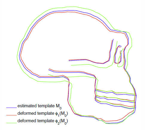
Establishing correspondence points across a set of biomedical shapes is an important technology for a variety of applications that rely on statistical analysis of individual subjects and populations. The inherent complexity (e.g. cortical surface shapes) and variability (e.g. cardiac chambers) evident in many biomedical shapes introduce significant challenges in finding a useful set of dense correspondences. Application specific strategies, such as registration of simplified (e.g. inflated or smoothed) surfaces or relying on manually placed landmarks, provide some improvement but suffer from limitations including increased computational complexity and ambiguity in landmark placement. This paper proposes a method for dense point correspondence on shape ensembles using geodesic distances to a priori landmarks as features. A novel set of numerical techniques for fast computation of geodesic distances to point sets is used to extract these features. The proposed method minimizes the ensemble entropy based on these features, resulting in isometry invariant correspondences in a very general, flexible framework.

D.J. Dosdall, R. Ranjan, K. Higuchi, E. Kholmovski, N. Angel, L. Li, R.S. Macleod, L. Norlund, A. Olsen, C.J. Davies, N.F. Marrouche.
“Chronic atrial fibrillation causes left ventricular dysfunction in dogs but not goats: experience with dogs, goats, and pigs,” In American Journal of Physiology: Heart and Circulatory Physiology, Vol. 305, No. 5, pp. H725--H731. September, 2013.
DOI: 10.1152/ajpheart.00440.2013
PubMed ID: 23812387
PubMed Central ID: PMC4116536
Structural remodeling in chronic atrial fibrillation (AF) occurs over weeks to months. To study the electrophysiological, structural, and functional changes that occur in chronic AF, the selection of the best animal model is critical. AF was induced by rapid atrial pacing (50-Hz stimulation every other second) in pigs (n = 4), dogs (n = 8), and goats (n = 9). Animals underwent MRIs at baseline and 6 mo to evaluate left ventricular (LV) ejection fraction (EF). Dogs were given metoprolol (50-100 mg po bid) and digoxin (0.0625-0.125 mg po bid) to limit the ventricular response rate to ot appropriate for chronic rapid atrial pacing-induced AF studies. Rate-controlled chronic AF in the dog model developed HF and LV fibrosis, whereas the goat model developed only atrial fibrosis without ventricular dysfunction and fibrosis. Both the dog and goat models are representative of segments of the patient population with chronic AF.
Keywords: animal models, chronic atrial fibrillation, fibrosis, heart failure, rapid atrial pacing
S. Durrleman, X. Pennec, A. Trouvé, J. Braga, G. Gerig, N. Ayache.
“Toward a comprehensive framework for the spatiotemporal statistical analysis of longitudinal shape data,” In International Journal of Computer Vision (IJCV), Vol. 103, No. 1, pp. 22--59. September, 2013.
DOI: 10.1007/s11263-012-0592-x
This paper proposes an original approach for the statistical analysis of longitudinal shape data. The proposed method allows the characterization of typical growth patterns and subject-specific shape changes in repeated time-series observations of several subjects. This can be seen as the extension of usual longitudinal statistics of scalar measurements to high-dimensional shape or image data.
The method is based on the estimation of continuous subject-specific growth trajectories and the comparison of such temporal shape changes across subjects. Differences between growth trajectories are decomposed into morphological deformations, which account for shape changes independent of the time, and time warps, which account for different rates of shape changes over time.
Given a longitudinal shape data set, we estimate a mean growth scenario representative of the population, and the variations of this scenario both in terms of shape changes and in terms of change in growth speed. Then, intrinsic statistics are derived in the space of spatiotemporal deformations, which characterize the typical variations in shape and in growth speed within the studied population. They can be used to detect systematic developmental delays across subjects.
In the context of neuroscience, we apply this method to analyze the differences in the growth of the hippocampus in children diagnosed with autism, developmental delays and in controls. Result suggest that group differences may be better characterized by a different speed of maturation rather than shape differences at a given age. In the context of anthropology, we assess the differences in the typical growth of the endocranium between chimpanzees and bonobos. We take advantage of this study to show the robustness of the method with respect to change of parameters and perturbation of the age estimates.
S. Durrleman, S. Allassonnière, S. Joshi.
“Sparse adaptive parameterization of variability in image ensembles,” In International Journal of Computer Vision (IJCV), Vol. 101, No. 1, pp. 161--183. 2013.
DOI: 10.1007/s11263-012-0556-1
L.T. Edgar, S.C. Sibole, C.J. Underwood, J.E. Guilkey, J.A. Weiss.
“A computational model of in vitro angiogenesis based on extracellular matrix fiber orientation,” In Computer Methods in Biomechanical and Biomedical Engineering, Vol. 16, No. 7, pp. 790--801. 2013.
DOI: 10.1080/10255842.2012.662678
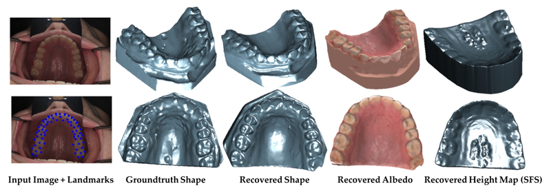
S. Elhabian, A. Farag, D. Tasman, W. Aboelmaaty, A. Farman.
“Clinical Crowns Shape Reconstruction - An Image-based Approach,” In Proceedings of the 2013 IEEE 10th International Symposium on Biomedical Imaging (ISBI), pp. 93--96. 2013.
DOI: 10.1109/ISBI.2013.6556420
J.T. Elison, J.J. Wolff, D.C. Heimer, S.J. Paterson, H. Gu, M. Styner, G. Gerig, J. Piven, the IBIS Network.
“Frontolimbic neural circuitry at 6 months predicts individual differences in joint attention at 9 months,” In Developmental Science, Vol. 16, No. 2, Wiley-Blackwell, pp. 186--197. 2013.
DOI: 10.1111/desc.12015
PubMed Central ID: PMC3582040

J.T. Elison, S.J. Paterson, J.J. Wolff, J.S. Reznick, N.J. Sasson, H. Gu, K.N. Botteron, S.R. Dager, A.M. Estes, A.C. Evans, G. Gerig, H.C. Hazlett, R.T. Schultz, M. Styner, L. Zwaigenbaum, J. Piven for the IBIS Network.
“White Matter Microstructure and Atypical Visual Orienting in 7 Month-Olds at Risk for Autism,” In American Journal of Psychiatry, Vol. AJP-12-09-1150.R2, March, 2013.
DOI: 10.1176/appi.ajp.2012.12091150
PubMed ID: 23511344
Objective: To determine whether specific patterns of oculomotor functioning and visual orienting characterize 7 month-old infants later classified with an autism spectrum disorder (ASD) and to identify the neural correlates of these behaviors.
Method: Ninety-seven infants contributed data to the current study (16 high-familial risk infants later classified with an ASD, 40 high-familial risk infants not meeting ASD criteria (high-risk-negative), and 41 low-risk infants). All infants completed an eye tracking task at 7 months and a clinical assessment at 25 months; diffusion weighted imaging data was acquired on 84 infants at 7 months. Primary outcome measures included average saccadic reaction time in a visually guided saccade procedure and radial diffusivity (an index of white matter organization) in fiber tracts that included corticospinal pathways and the splenium and genu of the corpus callosum.
Results: Visual orienting latencies were increased in seven-month-old infants who later express ASD symptoms at 25 months when compared with both high-risk-negative infants (p = 0.012, d = 0.73) and low-risk infants (p = 0.032, d = 0.71). Visual orienting latencies were uniquely associated with the microstructural organization of the splenium of the corpus callosum in low-risk infants, but this association was not apparent in infants later classified with ASD.
Conclusions: Flexibly and efficiently orienting to salient information in the environment is critical for subsequent cognitive and social-cognitive development. Atypical visual orienting may represent an earlyemerging prodromal feature of ASD, and abnormal functional specialization of posterior cortical circuits directly informs a novel model of ASD pathogenesis.
B. Erem, J. Coll-Font, R.M. Orellana, P. Stovicek, D.H. Brooks, R.S. MacLeod.
“Noninvasive reconstruction of potentials on endocardial surface from body surface potentials and CT imaging of partial torso,” In Journal of Electrocardiology, Vol. 46, No. 4, pp. e28. 2013.
DOI: 10.1016/j.jelectrocard.2013.05.104
B. Erem, R.M. Orellana, P. Stovicek, D.H. Brooks, R.S. MacLeod.
“Improved averaging of multi-lead ECGs and electrograms,” In Journal of Electrocardiology, Vol. 46, No. 4, Elsevier, pp. e28. July, 2013.
DOI: 10.1016/j.jelectrocard.2013.05.103
T. Etiene, D. Jonsson, T. Ropinski, C. Scheidegger, J. Comba, L. Gustavo Nonato, R.M. Kirby, A. Ynnerman, C.T. Silva.
“Verifying Volume Rendering Using Discretization Error Analysis,” SCI Technical Report, No. UUSCI-2013-001, SCI Institute, University of Utah, 2013.
Keywords: discretization errors, volume rendering, verifiable visualization
K. Fakhar, E. Hastings, C.R. Butson, K.D. Foote, P. Zeilman, M.S. Okun.
“Management of deep brain stimulator battery failure: battery estimators, charge density, and importance of clinical symptoms,” In PloS One, Vol. 8, No. 3, pp. e58665. January, 2013.
ISSN: 1932-6203
DOI: 10.1371/journal.pone.0058665
PubMed ID: 23536810
We aimed in this investigation to study deep brain stimulation (DBS) battery drain with special attention directed toward patient symptoms prior to and following battery replacement.
M. Farzinfar, Y. Li, A.R. Verde, I. Oguz, G. Gerig, M.A. Styner.
“DTI Quality Control Assessment via Error Estimation From Monte Carlo Simulations,” In Proceedings of SPIE 8669, Medical Imaging 2013: Image Processing, Vol. 8669, 2013.
DOI: 10.1117/12.2006925
PubMed ID: 23833547
PubMed Central ID: PMC3702180
M. Farzinfar, I. Oguz, R.G. Smith, A.R. Verde, C. Dietrich, A. Gupta, M.L. Escolar, J. Piven, S. Pujol, C. Vachet, S. Gouttard, G. Gerig, S. Dager, R.C. McKinstry, S. Paterson, A.C. Evans, M.A. Styner.
“Diffusion imaging quality control via entropy of principal direction distribution,” In NeuroImage, Vol. 82, pp. 1--12. 2013.
ISSN: 1053-8119
DOI: 10.1016/j.neuroimage.2013.05.022
Keywords: Diffusion magnetic resonance imaging, Diffusion tensor imaging, Quality assessment, Entropy
N. Farah, A. Zoubi, S. Matar, L. Golan, A. Marom, C.R. Butson, I. Brosh, S. Shoham.
“Holographically patterned activation using photo-absorber induced neural-thermal stimulation,” In Journal of Neural Engineering, Vol. 10, No. 5, pp. 056004. October, 2013.
ISSN: 1741-2560
DOI: 10.1088/1741-2560/10/5/056004

J. Fishbaugh, M.W. Prastawa, G. Gerig, S. Durrleman.
“Geodesic Shape Regression in the Framework of Currents,” In Proceedings of the International Conference on Information Processing in Medical Imaging (IPMI), Vol. 23, pp. 718--729. 2013.
PubMed ID: 24684012
PubMed Central ID: PMC4127488
J. Fishbaugh, M. Prastawa, G. Gerig, S. Durrleman.
“Geodesic image regression with a sparse parameterization of diffeomorphisms,” In Geometric Science of Information Lecture Notes in Computer Science (LNCS), In Proceedings of the Geometric Science of Information Conference (GSI), Vol. 8085, pp. 95--102. 2013.
Image regression allows for time-discrete imaging data to be modeled continuously, and is a crucial tool for conducting statistical analysis on longitudinal images. Geodesic models are particularly well suited for statistical analysis, as image evolution is fully characterized by a baseline image and initial momenta. However, existing geodesic image regression models are parameterized by a large number of initial momenta, equal to the number of image voxels. In this paper, we present a sparse geodesic image regression framework which greatly reduces the number of model parameters. We combine a control point formulation of deformations with a L1 penalty to select the most relevant subset of momenta. This way, the number of model parameters reflects the complexity of anatomical changes in time rather than the sampling of the image. We apply our method to both synthetic and real data and show that we can decrease the number of model parameters (from the number of voxels down to hundreds) with only minimal decrease in model accuracy. The reduction in model parameters has the potential to improve the power of ensuing statistical analysis, which faces the challenging problem of high dimensionality.
T. Fogal, A. Schiewe, J. Krüger.
“An Analysis of Scalable GPU-Based Ray-Guided Volume Rendering,” In 2013 IEEE Symposium on Large Data Analysis and Visualization (LDAV), 2013.
Volume rendering continues to be a critical method for analyzing large-scale scalar fields, in disciplines as diverse as biomedical engineering and computational fluid dynamics. Commodity desktop hardware has struggled to keep pace with data size increases, challenging modern visualization software to deliver responsive interactions for O(N3) algorithms such as volume rendering. We target the data type common in these domains: regularly-structured data.
In this work, we demonstrate that the major limitation of most volume rendering approaches is their inability to switch the data sampling rate (and thus data size) quickly. Using a volume renderer inspired by recent work, we demonstrate that the actual amount of visualizable data for a scene is typically bound considerably lower than the memory available on a commodity GPU. Our instrumented renderer is used to investigate design decisions typically swept under the rug in volume rendering literature. The renderer is freely available, with binaries for all major platforms as well as full source code, to encourage reproduction and comparison with future research.
Z. Fu, R.M. Kirby, R.T. Whitaker.
“A Fast Iterative Method for Solving the Eikonal Equation on Tetrahedral Domains,” In SIAM Journal on Scientific Computing, Vol. 35, No. 5, pp. C473--C494. 2013.
M. Gamell, I. Rodero, M. Parashar, J.C. Bennett, H. Kolla, J.H. Chen, P.-T. Bremer, A. Landge, A. Gyulassy, P. McCormick, Scott Pakin, Valerio Pascucci, Scott Klasky.
“Exploring Power Behaviors and Trade-offs of In-situ Data Analytics,” In Proceedings of the International Conference for High Performance Computing, Networking, Storage and Analysis, Association for Computing Machinery, 2013.
ISBN: 978-1-4503-2378-9
DOI: 10.1145/2503210.2503303
As scientific applications target exascale, challenges related to data and energy are becoming dominating concerns. For example, coupled simulation workflows are increasingly adopting in-situ data processing and analysis techniques to address costs and overheads due to data movement and I/O. However it is also critical to understand these overheads and associated trade-offs from an energy perspective. The goal of this paper is exploring data-related energy/performance trade-offs for end-to-end simulation workflows running at scale on current high-end computing systems. Specifically, this paper presents: (1) an analysis of the data-related behaviors of a combustion simulation workflow with an in-situ data analytics pipeline, running on the Titan system at ORNL; (2) a power model based on system power and data exchange patterns, which is empirically validated; and (3) the use of the model to characterize the energy behavior of the workflow and to explore energy/performance trade-offs on current as well as emerging systems.
Keywords: SDAV