Software Tools for Image Based Modeling, Simulation, and Visualization
| Organizers: | Rob MacLeod, PhD - University of Utah Dana Brooks, PhD - Northeastern University |
August 28, 2012 (Tuesday), 8:30 am - 5:30 pm.
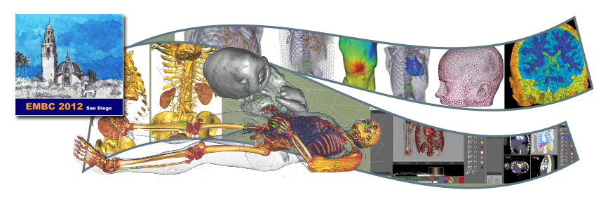
The goal of this tutorial is to introduce participants to a suite of software tools for image-based modeling, simulation, and visualization developed by the NIH/NIGMS Center for Integrative Biomedical Computing (CIBC). This portable flexible collection of interactive tools was designed in particular to support the development of subject specific, image based geometric models for simulation of bioelectric fields, but suite as well as its individual components have been applied to a wider set of problems. The tools in the suite are: ImageVis3D, for visualization of large scale data; Seg3D, for general purpose user-guided image segmentation; BioMesh3D, a set of utilities for creating surface and volume meshes from segmented image data; map3d, for visualization of surface based maps from multichannel time signals; and SCIRun, a comprehensive problem solving environment that integrates many of the capabilities of an entire image based modeling pipeline. The tutorial will be a mix of didactic presentations on the component steps of image based modeling, simulation, and visualization; hands on practice with the software, and cases studies on real world applications.
We will provide participants with the software and test data sets and encourage participants to bring their laptop computers ,and if relevant their own data as well. CIBC staff and developers will be on hand to help participants learn the programs, port their data, and generate useful results. We especially encourage participation by students, post docs, and technical users and software developers.
The tools in the suite include:
- Seg3D, for general purpose user-guided image segmentation;
- ImageVis3D, for visualization of large scale data;
- map3d, for visualization of surface based maps from multichannel time signals
- BioMesh3D, a set of utilities for creating surface and volume meshes from segmented image data;
- SCIRun, a comprehensive problem solving environment that integrates many of the capabilities of an entire image based modeling pipeline.
The tutorial will be a mix of didactic presentations on the components of image based modeling, simulation, and visualization; hands on practice with the software; and cases studies of real world applications. We will provide participants with the software and test data sets and encourage participants to bring their laptop computers, and, if relevant, their own data. CIBC members and developers will be on hand to help participants learn the programs, port their data, and generate useful results. We especially encourage participation by students, post docs, and technical users and software developers.
Intended audience
Scientists and engineers with an interest in applications of image processing, modeling, simulation, and visualization to their biomedical research.The goal of this tutorial is to introduce participants to a suite of software tools for image-based modeling, simulation, and visualization developed by the Center for Integrative Biomedical Computing (CIBC), an NIH-supported Biomedical Technology Research Center.
This portable flexible collection of interactive tools was designed to support the development of subject-specific, image-based geometric models for simulation of bioelectric fields. The tools, individually or as a suite, have also been applied to a wider set of problems.
Attendees will:
- learn about the major features of these software packages through presentations and case studies
- have an opportunity for hands-on instruction in our software, in laboratory sessions built into the workshop schedule
- interact with other users and technical personnel from our Center
- be able to try the tools on their own data with help from CIBC members
CIBC members and developers will be on hand to help participants learn the programs, port their data, and generate useful results.
We especially encourage participation by students, post docs, and technical users and software developers, as well as principal investigators.
Software Requirements
Graphics cards must support OpenGL 2.0 or greater. (not available on older Intel embedded graphics cards)
Windows: XP or better
OS X: 10.5 -10.7
SCIRun is not currently supported on OS X 10.8.
Installing platform-specific packages may be required.
Windows: Windows XP, Windows Vista, or Windows 7, Strongly recommended 64bit operating system
OS X: Mac OS X 10.5 - 10.8, 64-bit only
The Seg3D2 installer is not currently signed. The unsigned installer can be run on OS X 10.8 by control-clicking (or right-clicking if 2 button mouse support is enabled) on the installer package and selecting open.
CPU: minimum Core Duo or higher, recommended i5 or i7
Memory: minimum 4Gb, recommended 8Gb or more
Graphics Driver: minimum OpenGL 2.0 or higher (Not available on older Intel embedded graphics cards)
Graphics memory: 128, recommended 256Mb or more
Graphics cards must support OpenGL 2.0 or greater.
Python (2or 3) is required to run the BioMesh3D pipeline.
Multicore processor is strongly recommended.
Windows: Vista or 7
OS X: 10.5, 10.6
Installing platform-specific packages may be required.
BioMesh3D comes packaged with SCIRun.

Graphics cards must support OpenGL 2.0 or greater.
Windows: Windows XP or later
OS X: 10.7 or 10.8
Hardware, GPU: NVIDIA GeForce 9 series or better (i.e. GeForce 300M or later), or ATI/AMD Radeon HD 2400 series or better
Graphics cards must support OpenGL 2.0 or better.
Windows: Windows XP, Vista
OS X: 10.5 or greater
Required: 1.5 GHz CPU, 1GB RAM, 10GB free disk space, 64MB video RAM
Recommended: Dual-core 2 GHz CPU, 2GB RAM, 10GB free disk space, 128MB video RAM

Graphics cards must support OpenGL 2.0 or greater. (not available on older Intel embedded graphics cards)
Windows: XP or better
OS X: 10.5 -10.7
Installing platform-specific packages may be required.
Organizers and Speakers
 Rob MacLeod was trained in physics, electrical engineering, and physiology & biophysics and is an associate professor of Bioengineering and Medicine at the University of Utah. He is an associate director of the Scientific Computing and Imaging (SCI) Institute and the Nora Eccles Harrison Cardiovascular Research and Training Institute (CVRTI). He is an associate chairman and director of the undergraduate program in Biomedical Engineering and co-founder of the Center for Arrhythmia Research and Management (CARMA). His research interests include computational electrocardiography, experimental investigation and clinical detection of cardiac ischemia, cardiac arrhythmias, both atrial and ventricular, and defibrillation.
  Dana Brooks is a Professor in the Electrical and Computer Engineering Department at Northeastern University, the principal investigator of the BioMedical Imaging and Signal Processing Lab, an Associate Director of the Communications and Digital Signal Processing (CDSP) Center for Research and Graduate Education, and a researcher in the NSF-funded Gordon Center for Subsurface Sensing and Imaging Systems at NEU. Dr. Brooks is a member of the CIBC and his research interests include all areas of biomedical and biological signal and image processing, image based modeling, and especially forward and inverse problems in bioelectricity.
Speakers
 Tom Fogal, M.S. received his Computer Science degree from the University of New Hampshire (UNH). While an undergraduate he developed tools for visualizing mangetohydrodynamics data. As a graduate student, his interest in parallel rendering was cultured at UNH, Lawrence Livermore, and Oak Ridge national laboratories. Tom is the lead developer of ImageVis3D and the Tuvok imaging engine that drives it. At the CIBC, Tom also interacts directly with biomedical scientists and helps them learn and use the software as well as provides necessary interfaces to specific data types.
 Ayla Khan received her Bachelor of Computer Science in 2002 from Carleton University (Ottawa). As a developer for the CIBC, Ayla is the principal SCIRun developer and has also worked on other CIBC software products including Seg3D and BioMesh3D. Ayla is also working on her MS in the University of Utah's Computational Engineering and Science program.
 Moritz Dannhauer is a post-doctoral research associate at the Scientific Computing and Imaging (SCI) institute. Moritz is working on scientific problems concerning bioelectromagnetic field modeling
(Finite Element Method, Transcranial Direct Current Stimulation, etc.) and reconstruction methods for bioelectromagnetic sources.
 James Hughes received his B.S. in Mathematics from the University of Utah in 2011 and is working on ImageVis3D and other related CIBC projects. His research interests are: Simulation, Computer Graphics, Automated Reasoning and Control Theory.
 Manasi Datar is a Graduate Student working on statistical analysis of shape ensembles. More specifically, in characterizing the variability existing in such populations using particle correspondence models.
 Seyhmus Güler received his Bachelor's degree in Electrical and Electronics Engineering from Bilkent University(Turkey). As a graduate student in Northeastern University, Seyhmus is working on Current Optimization in Transcranial Direct Current Stimulation (tDCS). His research interests also include Finite Element Modelling, Forward and Inverse Problems in tDCS.
 Elizabeth Jurrus is currently serving a joint appointment for Pacific Northwest National Laboratory at the Scientific Computing and Imaging (SCI) Institute at the University of Utah. Her role at SCI includes technical manager for the NIH/NIGMS Center for Integrative Biomedical Computing, where she helps manages the development of open source tools for biomedical image-based modeling, biomedical simulation and estimation, and the visualization of biomedical data. Elizabeth also works as a liaison between the National Center for Microscopy and Imaging Research at UCSD and the image processing group at the SCI Institute to develop unique algorithms and tools for machine learning and image segmentation of neural processes in electron microscopy images. Current research interests focus on developing machine learning techniques for feature detection and the automated analysis of image data.