


Image Analysis
SCI's imaging work addresses fundamental questions in 2D and 3D image processing, including filtering, segmentation, surface reconstruction, and shape analysis. In low-level image processing, this effort has produce new nonparametric methods for modeling image statistics, which have resulted in better algorithms for denoising and reconstruction. Work with particle systems has led to new methods for visualizing and analyzing 3D surfaces. Our work in image processing also includes applications of advanced computing to 3D images, which has resulted in new parallel algorithms and real-time implementations on graphics processing units (GPUs). Application areas include medical image analysis, biological image processing, defense, environmental monitoring, and oil and gas.
Ross Whitaker
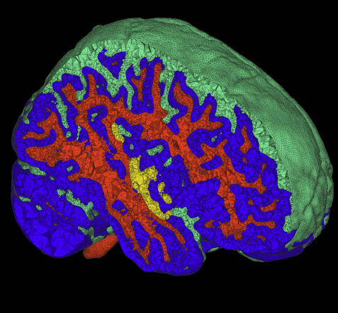
Segmentation

Chris Johnson
Diffusion Tensor Analysis
Funded Research Projects:
Publications in Image Analysis:
  Non-uniform Illumination Correction in Transmission Electron Microscopy T. Tasdizen , E. Jurrus, R. T. Whitaker. In MICCAI Workshop on Microscopic Image Analysis with Applications in Biology, 2008. Transmission electron microscopy (TEM) provides resolutions on the order of a nanometer. Hence, it is a critical imaging modality for biomedical analysis at the sub-cellular level. One of the problems associated with TEM images is variations in brightness due to electron imaging defects or non-uniform support films and specimen staining. These variations render image processing operations such as segmentation more difficult. The correction requires estimation of the global illumination field. In this paper, we propose an automatic method for estimating the illumination field using only image intensity gradients. The closed-form solution is very fast to compute. |
Cerebral Hypometabolism Suggesting Frototemporal Dementia in an Alzheimer’s Disease Clinical Trial N. L. Foster, A.Y. Wang, T. Tasdizen, K. Chen, W. Jagust, R.A. Koeppe, E. Reiman, M.W. Weiner, S. Minoshima. In Neurology, Vol. 70, No. 11, pp. A103. 2008. |
|
Multimaterial Meshing of MRI Head Data for Bioelectric Field Simulations R.T. Whitaker, R.M. Kirby, J.G. Sinstra, M.D. Meyer. In Proceedings of the 17th International Meshing Roundtable, 2008. The problem of body fitting meshes that are both adaptive and geometrically accurate is important in a variety of biomedical applications in a multitude of clinical settings, including electrocardiology, neurology, and orthopedics. Adaptivity is necessary because of the combination of large-scale and smallscale structures (e.g. relatively small blood vessels spanning a human head). Geometric accuracy is important for several reasons. In some cases, such as computational fluid dynamics, the fine-scale structure of the fluid domain is important for qualitative and quantitative accuracy of the solutions. More generally, finite element approximations of elliptic problems with rough coefficients require increased spatial resolution normal to material boundaries [3]. The problem of constructing meshes from biomedical images is particularly difficult because of the complexity and irregularity of the structures, and thus tuning or correcting meshes by hand is quite difficult and time consuming. Many researchers and, indeed, commercial products simply subdivide the underlying hexahedral image grid and assign material properties to tetrahedra based on standard decomposition of each hexahedron into tetrahedra. This paper presents a small case study of the results of a recently developed method for multimaterial, tetrahedral meshing of biomedical volumes [6]. The method uses an iterative relaxation of surface point point positions that are constrained to subsets of the volume that correspond to boundaries between different materials. In this paper we briefly review the method and present results on a set of MRI head images for use in bioelectric field simulation and source localization. |
|
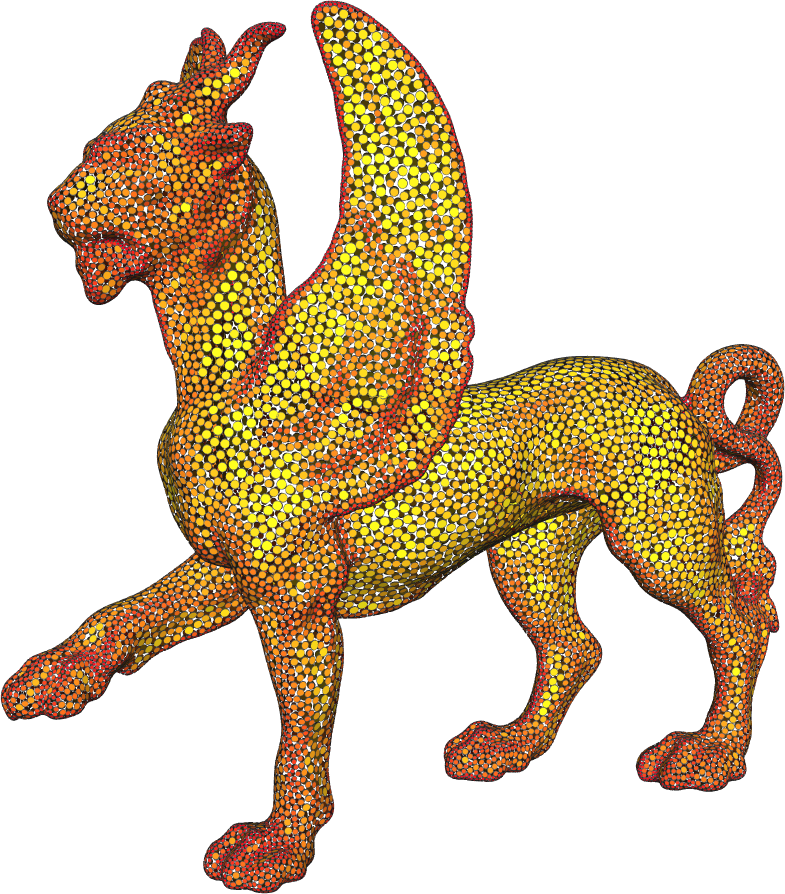
Dynamic Particle Systems for Adaptive Sampling of Implicit Surfaces M.D. Meyer. School of Computing, University of Utah, 2008. A ubiquitous requirement in many mathematical and computational problems is a set of well-placed point samples. For producing very even distributions of samples across complex surfaces, a dynamic particle system is a controllable mechanism that naturally accommodates strict sampling requirements. The systemfirst constrains particles to a surface, and then moves the particles across the surface until they are arranged in minimal energy configurations. Adaptivity is added into the system by scaling the distance between particles, causing higher densities of points around surface features. In this dissertation we explore and refine the dynamics of particle systems for generating efficient and adaptive point samples of implicit surfaces. Throughout this dissertation, we apply the adaptive particle system framework to several application areas. First, efficient visualizations of high-order finite element datasets are generated by developing adaptivity metrics of surfaces that exist in the presence of curvilinear coordinate transformation. Second, a framework is proposed that meets fundamental sampling constraints of Delaunay-based surface reconstruction algorithms. In meeting these constraints, the particle distributions produce nearly-regular, efficient isosurface tessellation that are geometrically and topologically accurate. And third, a novel analytic representation of material boundaries in multimaterial volume datasets is developed, as well as a set of projection operators, that allow for explicit sampling of nonmanifold material intersections. Using a tetrahedral labeling algorithm, the material intersections are extracted as watertight, nonmanifold meshes that are well-suited for simulations. |
| Particle-based Sampling and Meshing of Surfaces in Multimaterial Volumes M.D. Meyer, R.T. Whitaker, R.M. Kirby, C. Ledergerber, H. Pfister. In IEEE Transactions on Visualization and Computer Graphics, Vol. 14, No. 6, pp. 1539--1546. 2008. |
| An Optimal-Path Approach for Neural Circuit Reconstruction E. Jurrus, R.T. Whitaker, B. Jones, R. Marc, T. Tasdizen. In Proceedings of the 5th IEEE International Symposium on Biomedical Imaging: From Nano to Macro, pp. 1609--1612. 2008. PubMed ID: 19172170 |
Realizing the Potential of Positron Emission Tomography with 18F-Fluorodeoxyglucose to Improve the Treatment of Alzheimer N.L. Foster, A.Y. Wang, T. Tasdizen, P.T. Fletcher, J.M. Hoffman, R.A. Koeppe. In Journal of the Alzheimer, Vol. 4, No. 1, Suppl. 1, pp. S29--36. 2008. PubMed ID: 18631997 |
| Automatic Classification of Alzheimer N. Sadeghi, N.L. Foster, A.Y. Wang, S. Minoshima, A.P. Lieberman, T. Tasdizen. In Proceedings of IEEE International Symposium on Biomedical Imaging (ISBI 2008): From Nano to Macro, pp. 408--411. 2008. DOI: 10.1109/ISBI.2008.4541019 |
| Principal Components for Non-Local Means of Image Denoising T. Tasdizen. In Proceedings of the International Conference on Image Processing (ICIP 2008), pp. 1728--1731. 2008. PubMed ID: 19180227 |
| Fast Isosurface Extraction Methods for Large Image Data Sets Y. Livnat, S.G. Parker, C.R. Johnson. In Handbook of Medical Image Processing and Analysis, 2nd edition, Ch. 47, Note: (to appear), Edited by Isaac N. Bankman, Elsevier, pp. 801--816. 2008. |
| A Structural MRI Study of Human Brain Development from Birth to Two Years R.C. Knickmeyer, S. Gouttard, C. Kang, D. Evans, K. Wilber, K.J. Smith, R.M. Hamer, W. Lin, G. Gerig, J.H. Gilmore. In The Journal of Neuroscience, Vol. 28, No. 47, pp. 12176--12182. Nov, 2008. PubMed ID: 19020011 |
| CRA-NIH Computing Research Challenges in Biomedicine Workshop Recommendations D. Reed, C.R. Johnson. Note: Computing Research Association (CRA), 2007. |
| Robust Non-linear Dimensionality Reduction using Successive 1-Dimensional Laplacian Eigenmaps S. Gerber, T. Tasdizen, R.T. Whitaker. In Proceedings of the 2007 International Conference on Machine Learning (ICML), pp. 281--288. 2007. |
| Particle Systems for Efficient and Accurate Finite Element Visualization M.D. Meyer, B. Nelson, R.M. Kirby, R.T. Whitaker. In IEEE Transactions on Visualization and Computer Graphics, Vol. 13, No. 5, pp. 1015--1026. 2007. |
Anisotropic Curvature Motion for Structure Enhancing Smoothing of 3D MR Angiography Data O. Nemitz, T. Tasdizen, M. Rumpf, R.T. Whitaker. In Journal of Mathematical Imaging and Vision, Vol. 7, No. 3, pp. 217--229. 2007. DOI: 10.1007/s10851-006-0645-2 |
| Temporally Constrained Reconstruction of Dynamic Cardiac Perfusion MRI G. Adluru, S.P. Awate, T. Tasdizen, R.T. Whitaker, E.V.R. DiBella. In Magnetic Resonance in Medicine, Vol. 57, pp. 1027--1036. 2007. |
| Structure and Function of Microneuromas in Retinal Remodeling B.W. Jones, R.E. Marc, C.B. Watt, K. Kinardi, D. DeMill, J.H. Yang, T. Tasdizen, P. Koshevoy, E. Jurrus, R.T. Whitaker. In The Association for Research in Vision and Ophthalmology (ARVO) Conference, Note: (abstract), 2007. |
| Automatic Assembly of TEM Mosaics and Mosaic Stacks Using Phase Correlation SCI Institute Technical Report, P.A. Koshevoy, T. Tasdizen, R.T. Whitaker. No. UUSCI-2007-004, University of Utah, 2007. |
| Fast Isosurface Extraction Methods for Large Image Data Sets Y. Livnat, S.G. Parker, C.R. Johnson. In Handbook of Medical Imaging: Processing and Analysis, 2nd Edition, Ch. 44, Edited by Isaac Bankman, Academic Press, 2007. |
| Regional Gray Matter Growth, Sexual Dimorphism, and Cerebral Asymmetry in the Neonatal Brain J.H. Gilmore, W. Lin, M.W. Prastawa, C.B. Looney, Y.S.K. Vetsa, R.C. Knickmeyer, D.D. Evans, J.K. Smith, R.M. Hamer, J.A. Lieberman, G. Gerig. In Journal of Neuroscience, Vol. 27, No. 6, pp. 1255--1260. 2007. |